 Movie
Movie Controller

[English] 日本語
 Yorodumi
- PDB-2dbi: Crystal Structure of a Hypothetical Protein JW0805 from Escherich... -
+ Open data

- Basic information
| Entry | Database: PDB / ID: 2dbi | ||||||
|---|---|---|---|---|---|---|---|
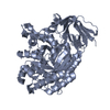
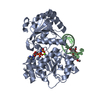
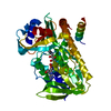
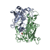
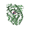
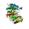
| Title | Crystal Structure of a Hypothetical Protein JW0805 from Escherichia coli | ||||||
 Components Components | Hypothetical protein ybiU | ||||||
 Keywords Keywords | Structural genomics / unknown function / alpha/beta structure / hypothetical protein / NPPSFA / National Project on Protein Structural and Functional Analyses / RIKEN Structural Genomics/Proteomics Initiative / RSGI | ||||||
| Function / homology | Gig2-like / Gig2-like / B-lactam Antibiotic, Isopenicillin N Synthase; Chain / Isopenicillin N synthase-like superfamily / Jelly Rolls / Sandwich / Mainly Beta / Uncharacterized protein YbiU Function and homology information Function and homology information | ||||||
| Biological species |  | ||||||
| Method |  X-RAY DIFFRACTION / SYNCHROTRON / MOLECULAR REPLACEMENT / Resolution: 2.05 Å X-RAY DIFFRACTION / SYNCHROTRON / MOLECULAR REPLACEMENT / Resolution: 2.05 Å | ||||||
 Authors Authors | Ihsanawati / Murayama, K. / Bessho, Y. / Shirouzu, M. / Yokoyama, S. / RIKEN Structural Genomics/Proteomics Initiative (RSGI) | ||||||
 Citation Citation | Journal: To be published Title: Crystal Structure of a Hypothetical Protein JW0805 from Escherichia coli Authors: Ihsanawati / Murayama, K. / Bessho, Y. / Shirouzu, M. / Yokoyama, S. | ||||||
| History |
|
- Structure visualization
| Structure viewer | Molecule: MolmilJmol/JSmol |
|---|
- Downloads & links
-Download
| PDBx/mmCIF format | 2dbi.cif.gz | 107.2 KB | Display | PDBx/mmCIF format |
|---|---|---|---|---|
| PDB format | pdb2dbi.ent.gz | 79.5 KB | Display | PDB format |
| PDBx/mmJSON format | 2dbi.json.gz | Tree view | PDBx/mmJSON format | |
| Others |  Other downloads Other downloads |
-Validation report
| Arichive directory | https://data.pdbj.org/pub/pdb/validation_reports/db/2dbiftp://data.pdbj.org/pub/pdb/validation_reports/db/2dbi | HTTPS FTP |
|---|
-Related structure data
| Related structure data | 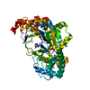 2dbnS S: Starting model for refinement |
|---|---|
| Similar structure data | |
| Other databases |


-Links
 PDBj
PDBj- Assembly
| Deposited unit | 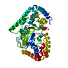
| ||||||||
|---|---|---|---|---|---|---|---|---|---|
| 1 |
| ||||||||
| Unit cell |
|
-Components
| #1: Protein | Mass: 51920.113 Da / Num. of mol.: 1 Source method: isolated from a genetically manipulated source Source: (gene. exp.) |
|---|---|
| #2: Water | ChemComp-HOH /  Mass: 18.015 Da / Num. of mol.: 617 / Source method: isolated from a natural source / Formula: H2O Mass: 18.015 Da / Num. of mol.: 617 / Source method: isolated from a natural source / Formula: H2O |
-Experimental details
-Experiment
| Experiment | Method: X-RAY DIFFRACTION / Number of used crystals: 1 |
|---|
- Sample preparation
| Crystal | Density Matthews: 2.38 Å3/Da / Density % sol: 48.28 % |
|---|---|
| Crystal grow | Temperature: 293 K / Method: vapor diffusion, hanging drop / pH: 5.5 Details: 0.1M bis-tris pH5.5, 25% PEG 3350, 0.2M NaCl, VAPOR DIFFUSION, HANGING DROP, temperature 293K |
-Data collection
| Diffraction | Mean temperature: 100 K |
|---|---|
| Diffraction source | Source: SYNCHROTRON / Site: SPring-8  / Beamline: BL26B2 / Wavelength: 1 Å / Beamline: BL26B2 / Wavelength: 1 Å |
| Detector | Type: RIGAKU JUPITER 210 / Detector: CCD / Date: Jul 20, 2005 |
| Radiation | Monochromator: Si / Protocol: SINGLE WAVELENGTH / Monochromatic (M) / Laue (L): M / Scattering type: x-ray |
| Radiation wavelength | Wavelength: 1 Å / Relative weight: 1 |
| Reflection | Resolution: 2→30 Å / Num. obs: 32097 / % possible obs: 93.8 % / Observed criterion σ(I): -3 / Biso Wilson estimate: 6.2 Å2 |
| Reflection shell | Resolution: 2→2.07 Å / % possible all: 36.6 |
- Processing
| Software |
| ||||||||||||||||||||
|---|---|---|---|---|---|---|---|---|---|---|---|---|---|---|---|---|---|---|---|---|---|
| Refinement | Method to determine structure: MOLECULAR REPLACEMENT Starting model: PDB ENTRY 2DBN Resolution: 2.05→30 Å / Rfactor Rfree error: 0.005 / Data cutoff high absF: 3570523.67 / Data cutoff low absF: 0 / Isotropic thermal model: RESTRAINED / Cross valid method: THROUGHOUT / σ(F): 0
| ||||||||||||||||||||
| Solvent computation | Solvent model: FLAT MODEL / Bsol: 56.7085 Å2 / ksol: 0.362091 e/Å3 | ||||||||||||||||||||
| Displacement parameters | Biso mean: 17.2 Å2
| ||||||||||||||||||||
| Refine analyze |
| ||||||||||||||||||||
| Refinement step | Cycle: LAST / Resolution: 2.05→30 Å
| ||||||||||||||||||||
| Refine LS restraints |
| ||||||||||||||||||||
| LS refinement shell | Resolution: 2.07→2.13 Å / Rfactor Rfree error: 0.017 / Total num. of bins used: 6
| ||||||||||||||||||||
| Xplor file |
|
