- PDB-2d68: Structure of the N-terminal domain of FOP (FGFR1OP) protein -
+
Open data
ID or keywords:
Loading...
-
Basic information
Entry
Database: PDB / ID: 2d68
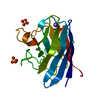
Title
Structure of the N-terminal domain of FOP (FGFR1OP) protein
Components
FOP
Keywords
CELL CYCLE / alpha helical bundle / dimer
Function / homology
Function and homology information
microtubule anchoring / Signaling by cytosolic FGFR1 fusion mutants / cell projection organization / protein tyrosine kinase inhibitor activity / negative regulation of protein kinase activity / Loss of Nlp from mitotic centrosomes / Loss of proteins required for interphase microtubule organization from the centrosome / Recruitment of mitotic centrosome proteins and complexes / Signaling by FGFR1 in disease / Recruitment of NuMA to mitotic centrosomes ...microtubule anchoring / Signaling by cytosolic FGFR1 fusion mutants / cell projection organization / protein tyrosine kinase inhibitor activity / negative regulation of protein kinase activity / Loss of Nlp from mitotic centrosomes / Loss of proteins required for interphase microtubule organization from the centrosome / Recruitment of mitotic centrosome proteins and complexes / Signaling by FGFR1 in disease / Recruitment of NuMA to mitotic centrosomes / centriole / Anchoring of the basal body to the plasma membrane / AURKA Activation by TPX2 / Regulation of PLK1 Activity at G2/M Transition / positive regulation of cell growth / ciliary basal body / positive regulation of cell migration / centrosome / positive regulation of cell population proliferation / protein kinase binding / perinuclear region of cytoplasm / protein homodimerization activity / nucleus / cytosol Similarity search - Function
In the structure databanks used in Yorodumi, some data are registered as the other names, "COVID-19 virus" and "2019-nCoV". Here are the details of the virus and the list of structure data.
Jan 31, 2019. EMDB accession codes are about to change! (news from PDBe EMDB page)
EMDB accession codes are about to change! (news from PDBe EMDB page)
The allocation of 4 digits for EMDB accession codes will soon come to an end. Whilst these codes will remain in use, new EMDB accession codes will include an additional digit and will expand incrementally as the available range of codes is exhausted. The current 4-digit format prefixed with “EMD-” (i.e. EMD-XXXX) will advance to a 5-digit format (i.e. EMD-XXXXX), and so on. It is currently estimated that the 4-digit codes will be depleted around Spring 2019, at which point the 5-digit format will come into force.
The EM Navigator/Yorodumi systems omit the EMD- prefix.
Related info.:Q: What is EMD? / ID/Accession-code notation in Yorodumi/EM Navigator
Yorodumi is a browser for structure data from EMDB, PDB, SASBDB, etc.
This page is also the successor to EM Navigator detail page, and also detail information page/front-end page for Omokage search.
The word "yorodu" (or yorozu) is an old Japanese word meaning "ten thousand". "mi" (miru) is to see.
Related info.:EMDB / PDB / SASBDB / Comparison of 3 databanks / Yorodumi Search / Aug 31, 2016. New EM Navigator & Yorodumi / Yorodumi Papers / Jmol/JSmol / Function and homology information / Changes in new EM Navigator and Yorodumi
 Movie
Movie Controller


 Open data
 Basic information
 Components
 Keywords
 Function and homology information
 Homo sapiens (human)
 X-RAY DIFFRACTION /
  Authors
 Citation
 Structure visualization
 Downloads & links
 Other downloads
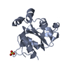


 PDBj
PDBj


 Assembly

 Mass: 18.015 Da / Num. of mol.: 200 / Source method: isolated from a natural source / Formula: H2O
 Sample preparation
 Processing