 Movie
Movie Controller

[English] 日本語
 Yorodumi
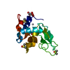
- PDB-2d4i: Monoclinic hen egg-white lysozyme crystallized at pH4.5 form heav... -
+ Open data

- Basic information
| Entry | Database: PDB / ID: 2d4i | ||||||
|---|---|---|---|---|---|---|---|
| Title | Monoclinic hen egg-white lysozyme crystallized at pH4.5 form heavy water solution | ||||||
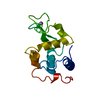
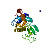
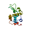
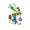
 Components Components | Lysozyme C | ||||||
 Keywords Keywords | HYDROLASE / phase transition / rigid-body motion / TLS analysis | ||||||
| Function / homology |  Function and homology information Function and homology informationLactose synthesis / Antimicrobial peptides / Neutrophil degranulation / beta-N-acetylglucosaminidase activity / cell wall macromolecule catabolic process / lysozyme / lysozyme activity / defense response to Gram-negative bacterium / killing of cells of another organism / defense response to Gram-positive bacterium ...Lactose synthesis / Antimicrobial peptides / Neutrophil degranulation / beta-N-acetylglucosaminidase activity / cell wall macromolecule catabolic process / lysozyme / lysozyme activity / defense response to Gram-negative bacterium / killing of cells of another organism / defense response to Gram-positive bacterium / defense response to bacterium / endoplasmic reticulum / extracellular space / identical protein binding / cytoplasm Similarity search - Function | ||||||
| Biological species |  | ||||||
| Method |  X-RAY DIFFRACTION / MOLECULAR REPLACEMENT / Resolution: 1.16 Å X-RAY DIFFRACTION / MOLECULAR REPLACEMENT / Resolution: 1.16 Å | ||||||
 Authors Authors | Harata, K. / Akiba, T. | ||||||
 Citation Citation | Journal: ACTA CRYSTALLOGR.,SECT.D / Year: 2006 Title: Structural phase transition of monoclinic crystals of hen egg-white lysozyme Authors: Harata, K. / Akiba, T. | ||||||
| History |
|
- Structure visualization
| Structure viewer | Molecule: MolmilJmol/JSmol |
|---|
- Downloads & links
-Download
| PDBx/mmCIF format | 2d4i.cif.gz | 127.9 KB | Display | PDBx/mmCIF format |
|---|---|---|---|---|
| PDB format | pdb2d4i.ent.gz | 100 KB | Display | PDB format |
| PDBx/mmJSON format | 2d4i.json.gz | Tree view | PDBx/mmJSON format | |
| Others |  Other downloads Other downloads |
-Validation report
| Summary document | 2d4i_validation.pdf.gz | 435.8 KB | Display | wwPDB validaton report |
|---|---|---|---|---|
| Full document | 2d4i_full_validation.pdf.gz | 439.9 KB | Display | |
| Data in XML | 2d4i_validation.xml.gz | 15.3 KB | Display | |
| Data in CIF | 2d4i_validation.cif.gz | 21.2 KB | Display | |
| Arichive directory | https://data.pdbj.org/pub/pdb/validation_reports/d4/2d4iftp://data.pdbj.org/pub/pdb/validation_reports/d4/2d4i | HTTPS FTP |
-Related structure data
| Related structure data | 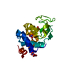 2d4jC 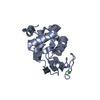 2d4kC 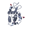 1lj4S C: citing same article ( S: Starting model for refinement |
|---|---|
| Similar structure data |

-Links
 PDBj
PDBj


- Assembly
| Deposited unit | 
| ||||||||
|---|---|---|---|---|---|---|---|---|---|
| 1 | 
| ||||||||
| 2 | 
| ||||||||
| Unit cell |
|
-Components
| #1: Protein | Mass: 14331.160 Da / Num. of mol.: 2 / Source method: isolated from a natural source / Source: (natural) #2: Chemical | ChemComp-NO3 /   Mass: 62.005 Da / Num. of mol.: 5 / Source method: obtained synthetically / Formula: NO3 Mass: 62.005 Da / Num. of mol.: 5 / Source method: obtained synthetically / Formula: NO3#3: Chemical | ChemComp-NA / |   Mass: 22.990 Da / Num. of mol.: 1 / Source method: obtained synthetically / Formula: Na Mass: 22.990 Da / Num. of mol.: 1 / Source method: obtained synthetically / Formula: Na#4: Chemical | ChemComp-DOD / |   Mass: 18.015 Da / Num. of mol.: 234 / Source method: isolated from a natural source / Formula: D2O Mass: 18.015 Da / Num. of mol.: 234 / Source method: isolated from a natural source / Formula: D2OHas protein modification | Y | |
|---|
-Experimental details
-Experiment
| Experiment | Method: X-RAY DIFFRACTION / Number of used crystals: 1 |
|---|
- Sample preparation
| Crystal | Density Matthews: 1.81 Å3/Da / Density % sol: 31.68 % |
|---|---|
| Crystal grow | Temperature: 298 K / Method: batch method / pH: 4.5 Details: 1% protein, 2% sodium nitrate, D2O solution with pD 4.5 adjusted with 1M acetic acid, batch method, temperature 298K |
-Data collection
| Diffraction | Mean temperature: 290 K |
|---|---|
| Diffraction source | Source: ROTATING ANODE / Type: MACSCIENCE / Wavelength: 1.5418 Å |
| Detector | Type: BRUKER SMART 6000 / Detector: CCD / Date: May 7, 2004 / Details: Conforcal Max-Flux optics |
| Radiation | Monochromator: curved mirror / Protocol: SINGLE WAVELENGTH / Monochromatic (M) / Laue (L): M / Scattering type: x-ray |
| Radiation wavelength | Wavelength: 1.5418 Å / Relative weight: 1 |
| Reflection | Resolution: 1.13→17.2 Å / Num. all: 77767 / Num. obs: 68963 / % possible obs: 88.7 % / Observed criterion σ(F): 0 / Observed criterion σ(I): 0 / Redundancy: 2.39 % / Rmerge(I) obs: 0.067 |
| Reflection shell | Resolution: 1.13→1.17 Å / Rmerge(I) obs: 0.415 / % possible all: 57.3 |
- Processing
| Software |
| |||||||||||||||||||||
|---|---|---|---|---|---|---|---|---|---|---|---|---|---|---|---|---|---|---|---|---|---|---|
| Refinement | Method to determine structure: MOLECULAR REPLACEMENT Starting model: PDB ENTRY 1LJ4 Resolution: 1.16→17.2 Å / σ(F): 0 / Stereochemistry target values: SHELX97 default Details: The author asigned the Rfree reflections by the following SHELXL97 input command line L.S. 5 -20 which indicates 5 cycles of least-squares calculation and ignore every 20th reflections (5%) ...Details: The author asigned the Rfree reflections by the following SHELXL97 input command line L.S. 5 -20 which indicates 5 cycles of least-squares calculation and ignore every 20th reflections (5%) in the refinement for the Rfree calculation
| |||||||||||||||||||||
| Refinement step | Cycle: LAST / Resolution: 1.16→17.2 Å
| |||||||||||||||||||||
| LS refinement shell | Resolution: 1.13→1.22 Å / Rfactor Rwork: 0.376 |
