- PDB-2d31: Crystal structure of disulfide-linked HLA-G dimer -
+
Open data
ID or keywords:
Loading...
-
Basic information
Entry
Database: PDB / ID: 2d31
Title
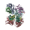
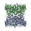
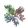
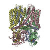
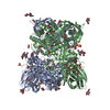
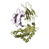
Crystal structure of disulfide-linked HLA-G dimer
Components
9-mer peptide from Histone H2A
Beta-2-microglobulin
HLA class I histocompatibility antigen, alpha chain G
Keywords
IMMUNE SYSTEM/CELL CYCLE / MHC class I / IMMUNE SYSTEM-CELL CYCLE COMPLEX
Function / homology
Function and homology information
peripheral B cell tolerance induction / positive regulation of tolerance induction / negative regulation of dendritic cell differentiation / positive regulation of natural killer cell cytokine production / positive regulation of T cell tolerance induction / immune response-inhibiting cell surface receptor signaling pathway / negative regulation of T cell mediated cytotoxicity / negative regulation of immune response / negative regulation of G0 to G1 transition / cis-Golgi network membrane ...peripheral B cell tolerance induction / positive regulation of tolerance induction / negative regulation of dendritic cell differentiation / positive regulation of natural killer cell cytokine production / positive regulation of T cell tolerance induction / immune response-inhibiting cell surface receptor signaling pathway / negative regulation of T cell mediated cytotoxicity / negative regulation of immune response / negative regulation of G0 to G1 transition / cis-Golgi network membrane / positive regulation of macrophage cytokine production / protein localization to site of double-strand break / negative regulation of natural killer cell mediated cytotoxicity / XY body / filopodium membrane / chromatin-protein adaptor activity / positive regulation of regulatory T cell differentiation / response to ionizing radiation / protein homotrimerization / CD8 receptor binding / protection from natural killer cell mediated cytotoxicity / positive regulation of endothelial cell apoptotic process / negative regulation of T cell proliferation / antigen processing and presentation of endogenous peptide antigen via MHC class Ib / antigen processing and presentation of endogenous peptide antigen via MHC class I via ER pathway, TAP-independent / positive regulation of interleukin-12 production / cellular defense response / site of DNA damage / Replacement of protamines by nucleosomes in the male pronucleus / Packaging Of Telomere Ends / DNA damage checkpoint signaling / male germ cell nucleus / Regulation of PD-L1(CD274) transcription / Recognition and association of DNA glycosylase with site containing an affected purine / Cleavage of the damaged purine / negative regulation of angiogenesis / Deposition of new CENPA-containing nucleosomes at the centromere / Recognition and association of DNA glycosylase with site containing an affected pyrimidine / Cleavage of the damaged pyrimidine / early endosome lumen / Nef mediated downregulation of MHC class I complex cell surface expression / DAP12 interactions / RNA Polymerase I Promoter Opening / Inhibition of DNA recombination at telomere / Assembly of the ORC complex at the origin of replication / negative regulation of phosphatidylinositol 3-kinase/protein kinase B signal transduction / Meiotic synapsis / T cell mediated cytotoxicity / positive regulation of DNA repair / Regulation of endogenous retroelements by the Human Silencing Hub (HUSH) complex / DNA methylation / Condensation of Prophase Chromosomes / condensed nuclear chromosome / Endosomal/Vacuolar pathway / Chromatin modifications during the maternal to zygotic transition (MZT) / SIRT1 negatively regulates rRNA expression / ERCC6 (CSB) and EHMT2 (G9a) positively regulate rRNA expression / replication fork / PRC2 methylates histones and DNA / Regulation of endogenous retroelements by KRAB-ZFP proteins / Antigen Presentation: Folding, assembly and peptide loading of class I MHC / Defective pyroptosis / lumenal side of endoplasmic reticulum membrane / antigen processing and presentation of exogenous protein antigen via MHC class Ib, TAP-dependent / regulation of iron ion transport / cellular response to iron(III) ion / negative regulation of iron ion transport / negative regulation of forebrain neuron differentiation / response to molecule of bacterial origin / regulation of erythrocyte differentiation / Regulation of endogenous retroelements by Piwi-interacting RNAs (piRNAs) / peptide antigen assembly with MHC class I protein complex / ER to Golgi transport vesicle membrane / RNA Polymerase I Promoter Escape / Nonhomologous End-Joining (NHEJ) / Transcriptional regulation by small RNAs / HFE-transferrin receptor complex / MHC class I peptide loading complex / transferrin transport / negative regulation of receptor-mediated endocytosis / cellular response to iron ion / positive regulation of T cell cytokine production / antigen processing and presentation of endogenous peptide antigen via MHC class I / Formation of the beta-catenin:TCF transactivating complex / meiotic cell cycle / Activated PKN1 stimulates transcription of AR (androgen receptor) regulated genes KLK2 and KLK3 / MHC class I protein complex / RUNX1 regulates genes involved in megakaryocyte differentiation and platelet function / multicellular organismal-level iron ion homeostasis / peptide antigen assembly with MHC class II protein complex / positive regulation of T cell mediated cytotoxicity / negative regulation of epithelial cell proliferation / cellular response to nicotine / MHC class II protein complex / positive regulation of receptor-mediated endocytosis / negative regulation of neurogenesis / Negative Regulation of CDH1 Gene Transcription / NoRC negatively regulates rRNA expression / G2/M DNA damage checkpoint / B-WICH complex positively regulates rRNA expression Similarity search - Function
MHC class I-like antigen recognition-like / Murine Class I Major Histocompatibility Complex, H2-DB; Chain A, domain 1 / MHC class I alpha chain, alpha1 alpha2 domains / Class I Histocompatibility antigen, domains alpha 1 and 2 / Beta-2-Microglobulin / : / MHC class I-like antigen recognition-like / MHC class I-like antigen recognition-like superfamily / Histone H2A conserved site / Histone H2A signature. ...MHC class I-like antigen recognition-like / Murine Class I Major Histocompatibility Complex, H2-DB; Chain A, domain 1 / MHC class I alpha chain, alpha1 alpha2 domains / Class I Histocompatibility antigen, domains alpha 1 and 2 / Beta-2-Microglobulin / : / MHC class I-like antigen recognition-like / MHC class I-like antigen recognition-like superfamily / Histone H2A conserved site / Histone H2A signature. / Histone H2A, C-terminal domain / C-terminus of histone H2A / Histone 2A / Histone H2A / MHC classes I/II-like antigen recognition protein / : / Histone H2A/H2B/H3 / Core histone H2A/H2B/H3/H4 domain / Histone-fold / Immunoglobulin/major histocompatibility complex, conserved site / Immunoglobulins and major histocompatibility complex proteins signature. / Immunoglobulin C-Type / Immunoglobulin C1-set / Immunoglobulin C1-set domain / Ig-like domain profile. / Immunoglobulin-like domain / Immunoglobulin-like domain superfamily / Immunoglobulin-like fold / Immunoglobulins / Immunoglobulin-like / Sandwich / 2-Layer Sandwich / Mainly Beta / Alpha Beta Similarity search - Domain/homology
Histone H2AX / HLA class I histocompatibility antigen, alpha chain G / Beta-2-microglobulin Similarity search - Component
A: HLA class I histocompatibility antigen, alpha chain G B: Beta-2-microglobulin C: 9-mer peptide from Histone H2A D: HLA class I histocompatibility antigen, alpha chain G E: Beta-2-microglobulin F: 9-mer peptide from Histone H2A
In the structure databanks used in Yorodumi, some data are registered as the other names, "COVID-19 virus" and "2019-nCoV". Here are the details of the virus and the list of structure data.
Jan 31, 2019. EMDB accession codes are about to change! (news from PDBe EMDB page)
EMDB accession codes are about to change! (news from PDBe EMDB page)
The allocation of 4 digits for EMDB accession codes will soon come to an end. Whilst these codes will remain in use, new EMDB accession codes will include an additional digit and will expand incrementally as the available range of codes is exhausted. The current 4-digit format prefixed with “EMD-” (i.e. EMD-XXXX) will advance to a 5-digit format (i.e. EMD-XXXXX), and so on. It is currently estimated that the 4-digit codes will be depleted around Spring 2019, at which point the 5-digit format will come into force.
The EM Navigator/Yorodumi systems omit the EMD- prefix.
Related info.:Q: What is EMD? / ID/Accession-code notation in Yorodumi/EM Navigator
Yorodumi is a browser for structure data from EMDB, PDB, SASBDB, etc.
This page is also the successor to EM Navigator detail page, and also detail information page/front-end page for Omokage search.
The word "yorodu" (or yorozu) is an old Japanese word meaning "ten thousand". "mi" (miru) is to see.
Related info.:EMDB / PDB / SASBDB / Comparison of 3 databanks / Yorodumi Search / Aug 31, 2016. New EM Navigator & Yorodumi / Yorodumi Papers / Jmol/JSmol / Function and homology information / Changes in new EM Navigator and Yorodumi
 Movie
Movie Controller


 Open data
 Basic information
 Components
 Keywords
 Function and homology information
 Homo sapiens (human)
 X-RAY DIFFRACTION /
  Authors
 Citation
 Structure visualization
 Downloads & links
 Other downloads
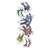

 PDBj
PDBj


 Assembly


 Sample preparation
 / Beamline: BL38B1 / Wavelength: 1 Å
 Processing