 Movie
Movie Controller

[English] 日本語
 Yorodumi
- PDB-2d1g: Structure of Francisella tularensis Acid Phosphatase A (AcpA) bou... -
+ Open data

- Basic information
| Entry | Database: PDB / ID: 2d1g | ||||||
|---|---|---|---|---|---|---|---|
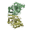
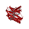
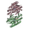
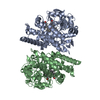
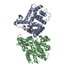
| Title | Structure of Francisella tularensis Acid Phosphatase A (AcpA) bound to orthovanadate | ||||||
 Components Components | acid phosphatase | ||||||
 Keywords Keywords | HYDROLASE / Francisella tularensis / Acid Phosphatase / AcpA / decavanadate / vanadate | ||||||
| Function / homology |  Function and homology information Function and homology information | ||||||
| Biological species |  Francisella tularensis subsp. novicida (bacteria) Francisella tularensis subsp. novicida (bacteria) | ||||||
| Method |  X-RAY DIFFRACTION / SYNCHROTRON / SAD / Resolution: 1.75 Å X-RAY DIFFRACTION / SYNCHROTRON / SAD / Resolution: 1.75 Å | ||||||
 Authors Authors | Felts, R.L. / Reilly, T.J. / Tanner, J.J. | ||||||
 Citation Citation | Journal: J.Biol.Chem. / Year: 2006 Title: Structure of Francisella tularensis AcpA: prototype of a unique superfamily of acid phosphatases and phospholipases C Authors: Felts, R.L. / Reilly, T.J. / Tanner, J.J. | ||||||
| History |
|
- Structure visualization
| Structure viewer | Molecule: MolmilJmol/JSmol |
|---|
- Downloads & links
-Download
| PDBx/mmCIF format | 2d1g.cif.gz | 209.7 KB | Display | PDBx/mmCIF format |
|---|---|---|---|---|
| PDB format | pdb2d1g.ent.gz | 166.9 KB | Display | PDB format |
| PDBx/mmJSON format | 2d1g.json.gz | Tree view | PDBx/mmJSON format | |
| Others |  Other downloads Other downloads |
-Validation report
| Arichive directory | https://data.pdbj.org/pub/pdb/validation_reports/d1/2d1gftp://data.pdbj.org/pub/pdb/validation_reports/d1/2d1g | HTTPS FTP |
|---|
-Related structure data
| Similar structure data |
|---|

-Links
 PDBj
PDBj
- Assembly
| Deposited unit | 
| ||||||||
|---|---|---|---|---|---|---|---|---|---|
| 1 |
| ||||||||
| Unit cell |
|
-Components
-Protein , 1 types, 2 molecules AB
| #1: Protein | Mass: 56338.754 Da / Num. of mol.: 2 Source method: isolated from a genetically manipulated source Source: (gene. exp.) Francisella tularensis subsp. novicida (bacteria)Species: Francisella tularensis / Strain: subsp. novicida / Gene: acpA / Plasmid: pET20b / Species (production host): Escherichia coli / Production host: References: UniProt: Q2A5P7, UniProt: A0Q436*PLUS, acid phosphatase |
|---|
-Non-polymers , 7 types, 559 molecules 


| #2: Chemical |  Mass: 114.939 Da / Num. of mol.: 2 / Source method: obtained synthetically / Formula: VO4 Mass: 114.939 Da / Num. of mol.: 2 / Source method: obtained synthetically / Formula: VO4#3: Chemical |  Num. of mol.: 2 / Source method: obtained synthetically Num. of mol.: 2 / Source method: obtained synthetically#4: Chemical | ChemComp-ETE / |  Mass: 208.252 Da / Num. of mol.: 1 / Source method: obtained synthetically / Formula: C9H20O5 Mass: 208.252 Da / Num. of mol.: 1 / Source method: obtained synthetically / Formula: C9H20O5#5: Chemical | ChemComp-ETX / |  Mass: 90.121 Da / Num. of mol.: 1 / Source method: obtained synthetically / Formula: C4H10O2 Mass: 90.121 Da / Num. of mol.: 1 / Source method: obtained synthetically / Formula: C4H10O2#6: Chemical | ChemComp-DVT / |  Mass: 957.398 Da / Num. of mol.: 1 / Source method: obtained synthetically / Formula: O28V10 Mass: 957.398 Da / Num. of mol.: 1 / Source method: obtained synthetically / Formula: O28V10#7: Chemical |  Mass: 150.173 Da / Num. of mol.: 2 / Source method: obtained synthetically / Formula: C6H14O4 Mass: 150.173 Da / Num. of mol.: 2 / Source method: obtained synthetically / Formula: C6H14O4#8: Water | ChemComp-HOH / | Mass: 18.015 Da / Num. of mol.: 550 / Source method: isolated from a natural source / Formula: H2O |
|---|
-Details
| Has protein modification | Y |
|---|
-Experimental details
-Experiment
| Experiment | Method: X-RAY DIFFRACTION / Number of used crystals: 2 |
|---|
- Sample preparation
| Crystal | Density Matthews: 2.2 Å3/Da / Density % sol: 43.9 % |
|---|---|
| Crystal grow | Temperature: 298 K / Method: vapor diffusion, sitting drop / pH: 6 Details: PEG 1500, pH 6, VAPOR DIFFUSION, SITTING DROP, temperature 298K |
-Data collection
| Diffraction |
| |||||||||||||||||||||||||||||||||||||||||||||||||||||||||||||||||||||||||||||
|---|---|---|---|---|---|---|---|---|---|---|---|---|---|---|---|---|---|---|---|---|---|---|---|---|---|---|---|---|---|---|---|---|---|---|---|---|---|---|---|---|---|---|---|---|---|---|---|---|---|---|---|---|---|---|---|---|---|---|---|---|---|---|---|---|---|---|---|---|---|---|---|---|---|---|---|---|---|---|
| Diffraction source |
| |||||||||||||||||||||||||||||||||||||||||||||||||||||||||||||||||||||||||||||
| Detector |
| |||||||||||||||||||||||||||||||||||||||||||||||||||||||||||||||||||||||||||||
| Radiation |
| |||||||||||||||||||||||||||||||||||||||||||||||||||||||||||||||||||||||||||||
| Radiation wavelength |
| |||||||||||||||||||||||||||||||||||||||||||||||||||||||||||||||||||||||||||||
| Reflection | Redundancy: 14.4 % / Number: 39861 / Rmerge(I) obs: 0.065 / Χ2: 0.985 / D res high: 2.39 Å / D res low: 50 Å / % possible obs: 99.9 | |||||||||||||||||||||||||||||||||||||||||||||||||||||||||||||||||||||||||||||
| Diffraction reflection shell |
| |||||||||||||||||||||||||||||||||||||||||||||||||||||||||||||||||||||||||||||
| Reflection | Resolution: 1.75→50 Å / Num. obs: 98137 / % possible obs: 97.2 % / Observed criterion σ(F): 2 / Observed criterion σ(I): 2 / Redundancy: 4.1 % / Rmerge(I) obs: 0.047 / Χ2: 1.015 | |||||||||||||||||||||||||||||||||||||||||||||||||||||||||||||||||||||||||||||
| Reflection shell | Resolution: 1.75→1.81 Å / % possible obs: 94.9 % / Redundancy: 4 % / Rmerge(I) obs: 0.357 / Num. measured obs: 9497 / Χ2: 0.975 |

-Phasing
| Phasing | Method: SAD |
|---|---|
| Phasing MAD | D res high: 1.75 Å / D res low: 44.23 Å / FOM acentric: 0.364 / FOM centric: 0.489 / Reflection acentric: 92746 / Reflection centric: 5082 |
| Phasing dm shell | Resolution: 1.75→50 Å / Delta phi final: 0.29 / FOM : 0.315 / Reflection: 98049 |
- Processing
| Software |
| ||||||||||||||||||||||||||||||||||||||||||||||||||||||||||||||||||||||||||||||||||||||||||||||||||||
|---|---|---|---|---|---|---|---|---|---|---|---|---|---|---|---|---|---|---|---|---|---|---|---|---|---|---|---|---|---|---|---|---|---|---|---|---|---|---|---|---|---|---|---|---|---|---|---|---|---|---|---|---|---|---|---|---|---|---|---|---|---|---|---|---|---|---|---|---|---|---|---|---|---|---|---|---|---|---|---|---|---|---|---|---|---|---|---|---|---|---|---|---|---|---|---|---|---|---|---|---|---|
| Refinement | Method to determine structure: SAD / Resolution: 1.75→44.28 Å / Cor.coef. Fo:Fc: 0.952 / Cor.coef. Fo:Fc free: 0.936 / SU B: 5.27 / SU ML: 0.09 / TLS residual ADP flag: LIKELY RESIDUAL / Cross valid method: THROUGHOUT / σ(F): 0 / ESU R: 0.13 / ESU R Free: 0.122 / Stereochemistry target values: MAXIMUM LIKELIHOOD
| ||||||||||||||||||||||||||||||||||||||||||||||||||||||||||||||||||||||||||||||||||||||||||||||||||||
| Solvent computation | Ion probe radii: 0.8 Å / Shrinkage radii: 0.8 Å / VDW probe radii: 1.2 Å / Solvent model: MASK | ||||||||||||||||||||||||||||||||||||||||||||||||||||||||||||||||||||||||||||||||||||||||||||||||||||
| Displacement parameters | Biso mean: 25.27 Å2
| ||||||||||||||||||||||||||||||||||||||||||||||||||||||||||||||||||||||||||||||||||||||||||||||||||||
| Refinement step | Cycle: LAST / Resolution: 1.75→44.28 Å
| ||||||||||||||||||||||||||||||||||||||||||||||||||||||||||||||||||||||||||||||||||||||||||||||||||||
| Refine LS restraints |
| ||||||||||||||||||||||||||||||||||||||||||||||||||||||||||||||||||||||||||||||||||||||||||||||||||||
| LS refinement shell | Resolution: 1.75→1.796 Å / Total num. of bins used: 20
| ||||||||||||||||||||||||||||||||||||||||||||||||||||||||||||||||||||||||||||||||||||||||||||||||||||
| Refinement TLS params. | Method: refined / Refine-ID: X-RAY DIFFRACTION
| ||||||||||||||||||||||||||||||||||||||||||||||||||||||||||||||||||||||||||||||||||||||||||||||||||||
| Refinement TLS group |
|
