 Movie
Movie Controller

[English] 日本語
 Yorodumi
- PDB-2cy0: Crystal Structure of Shikimate 5-Dehydrogenase (AroE) from Thermu... -
+ Open data

- Basic information
| Entry | Database: PDB / ID: 2cy0 | ||||||
|---|---|---|---|---|---|---|---|
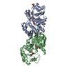
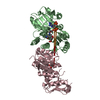
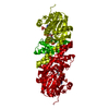
| Title | Crystal Structure of Shikimate 5-Dehydrogenase (AroE) from Thermus Thermophilus HB8 in complex with NADP | ||||||
 Components Components | shikimate 5-dehydrogenase | ||||||
 Keywords Keywords | OXIDOREDUCTASE / Shikimate / Cofactor / Dimer / Structural Genomics / NPPSFA / National Project on Protein Structural and Functional Analyses / RIKEN Structural Genomics/Proteomics Initiative / RSGI | ||||||
| Function / homology |  Function and homology information Function and homology informationshikimate dehydrogenase (NADP+) / shikimate 3-dehydrogenase (NADP+) activity / shikimate metabolic process / chorismate biosynthetic process / aromatic amino acid biosynthetic process / amino acid biosynthetic process / NADP binding / cytosol Similarity search - Function | ||||||
| Biological species |   Thermus thermophilus (bacteria) Thermus thermophilus (bacteria) | ||||||
| Method |  X-RAY DIFFRACTION / MOLECULAR REPLACEMENT / Resolution: 1.9 Å X-RAY DIFFRACTION / MOLECULAR REPLACEMENT / Resolution: 1.9 Å | ||||||
 Authors Authors | Bagautdinov, B. / Kunishima, N. / RIKEN Structural Genomics/Proteomics Initiative (RSGI) | ||||||
 Citation Citation | Journal: J.Mol.Biol. / Year: 2007 Title: Crystal Structures of Shikimate Dehydrogenase AroE from Thermus thermophilus HB8 and its Cofactor and Substrate Complexes: Insights into the Enzymatic Mechanism Authors: Bagautdinov, B. / Kunishima, N. | ||||||
| History |
|
- Structure visualization
| Structure viewer | Molecule: MolmilJmol/JSmol |
|---|
- Downloads & links
-Download
| PDBx/mmCIF format | 2cy0.cif.gz | 124.5 KB | Display | PDBx/mmCIF format |
|---|---|---|---|---|
| PDB format | pdb2cy0.ent.gz | 95.6 KB | Display | PDB format |
| PDBx/mmJSON format | 2cy0.json.gz | Tree view | PDBx/mmJSON format | |
| Others |  Other downloads Other downloads |
-Validation report
| Arichive directory | https://data.pdbj.org/pub/pdb/validation_reports/cy/2cy0ftp://data.pdbj.org/pub/pdb/validation_reports/cy/2cy0 | HTTPS FTP |
|---|
-Related structure data
| Related structure data |  1wxdSC  2d5cC  2ev9C S: Starting model for refinement C: citing same article ( |
|---|---|
| Similar structure data | |
| Other databases |


-Links
 PDBj
PDBj- Assembly
| Deposited unit | 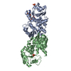
| ||||||||
|---|---|---|---|---|---|---|---|---|---|
| 1 |
| ||||||||
| Unit cell |
|
-Components
| #1: Protein | Mass: 28316.791 Da / Num. of mol.: 2 Source method: isolated from a genetically manipulated source Source: (gene. exp.) Thermus thermophilus (bacteria) / Strain: HB8 / Gene: AroE / Plasmid: pET 11a / Species (production host): Escherichia coli / Production host: References: UniProt: Q5SJF8, shikimate dehydrogenase (NADP+) #2: Chemical |   Mass: 96.063 Da / Num. of mol.: 2 / Source method: obtained synthetically / Formula: SO4 Mass: 96.063 Da / Num. of mol.: 2 / Source method: obtained synthetically / Formula: SO4#3: Chemical | ChemComp-NAP / |   Mass: 743.405 Da / Num. of mol.: 1 / Source method: obtained synthetically / Formula: C21H28N7O17P3 Mass: 743.405 Da / Num. of mol.: 1 / Source method: obtained synthetically / Formula: C21H28N7O17P3#4: Water | ChemComp-HOH / |  Mass: 18.015 Da / Num. of mol.: 516 / Source method: isolated from a natural source / Formula: H2O Mass: 18.015 Da / Num. of mol.: 516 / Source method: isolated from a natural source / Formula: H2O |
|---|
-Experimental details
-Experiment
| Experiment | Method: X-RAY DIFFRACTION / Number of used crystals: 1 |
|---|
- Sample preparation
| Crystal | Density Matthews: 2.22 Å3/Da / Density % sol: 44.6 % |
|---|---|
| Crystal grow | Temperature: 295 K / Method: microbatch / pH: 4.6 Details: Ammonium sulfate, Sodium Acetate trihydrate, PEG4000, pH 4.6, microbatch, temperature 295K |
-Data collection
| Diffraction | Mean temperature: 100 K |
|---|---|
| Diffraction source | Source: ROTATING ANODE / Type: RIGAKU FR-D / Wavelength: 1.5418 Å |
| Detector | Type: RIGAKU RAXIS IV / Detector: IMAGE PLATE / Date: Jan 12, 2005 / Details: mirrors |
| Radiation | Monochromator: GRAPHITE / Protocol: SINGLE WAVELENGTH / Monochromatic (M) / Laue (L): M / Scattering type: x-ray |
| Radiation wavelength | Wavelength: 1.5418 Å / Relative weight: 1 |
| Reflection | Resolution: 1.9→31.15 Å / Num. all: 39339 / Num. obs: 39285 / % possible obs: 96.3 % / Observed criterion σ(F): 0 / Observed criterion σ(I): 0 / Redundancy: 4.2 % / Biso Wilson estimate: 26.1 Å2 / Rmerge(I) obs: 0.052 |
| Reflection shell | Resolution: 1.9→1.97 Å / Redundancy: 2.8 % / Rmerge(I) obs: 0.468 / Mean I/σ(I) obs: 2.3 / Num. unique all: 3487 / % possible all: 87.5 |
- Processing
| Software |
| |||||||||||||||||||||||||
|---|---|---|---|---|---|---|---|---|---|---|---|---|---|---|---|---|---|---|---|---|---|---|---|---|---|---|
| Refinement | Method to determine structure: MOLECULAR REPLACEMENT Starting model: PDB ENTRY 1WXD Resolution: 1.9→31.15 Å / Isotropic thermal model: OVERALL / Cross valid method: THROUGHOUT / σ(F): 0 / σ(I): 0 / Stereochemistry target values: Engh & Huber
| |||||||||||||||||||||||||
| Displacement parameters | Biso mean: 36.6 Å2
| |||||||||||||||||||||||||
| Refine analyze |
| |||||||||||||||||||||||||
| Refinement step | Cycle: LAST / Resolution: 1.9→31.15 Å
| |||||||||||||||||||||||||
| Refine LS restraints |
|
