 Movie
Movie Controller

[English] 日本語
 Yorodumi
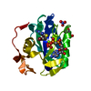
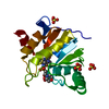
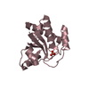
- PDB-2cwd: Crystal Structure of TT1001 protein from Thermus thermophilus HB8 -
+ Open data

- Basic information
| Entry | Database: PDB / ID: 2cwd | ||||||
|---|---|---|---|---|---|---|---|
| Title | Crystal Structure of TT1001 protein from Thermus thermophilus HB8 | ||||||
 Components Components | low molecular weight phosphotyrosine protein phosphatase | ||||||
 Keywords Keywords | HYDROLASE / tyrosine phosphatase / structural genomics / RIKEN Structural Genomics/Proteomics Initiative / RSGI / NPPSFA / National Project on Protein Structural and Functional Analyses | ||||||
| Function / homology |  Function and homology information Function and homology informationprotein-tyrosine-phosphatase / protein tyrosine phosphatase activity / metal ion binding Similarity search - Function | ||||||
| Biological species |   Thermus thermophilus (bacteria) Thermus thermophilus (bacteria) | ||||||
| Method |  X-RAY DIFFRACTION / SYNCHROTRON / MOLECULAR REPLACEMENT / Resolution: 1.9 Å X-RAY DIFFRACTION / SYNCHROTRON / MOLECULAR REPLACEMENT / Resolution: 1.9 Å | ||||||
 Authors Authors | Lokanath, N.K. / Terao, Y. / Kunishima, N. / RIKEN Structural Genomics/Proteomics Initiative (RSGI) | ||||||
 Citation Citation | Journal: To be Published Title: Crystal Structure of TT1001 protein from Thermus thermophilus HB8 Authors: Lokanath, N.K. / Terao, Y. / Kunishima, N. | ||||||
| History |
|
- Structure visualization
| Structure viewer | Molecule: MolmilJmol/JSmol |
|---|
- Downloads & links
-Download
| PDBx/mmCIF format | 2cwd.cif.gz | 140.1 KB | Display | PDBx/mmCIF format |
|---|---|---|---|---|
| PDB format | pdb2cwd.ent.gz | 109.7 KB | Display | PDB format |
| PDBx/mmJSON format | 2cwd.json.gz | Tree view | PDBx/mmJSON format | |
| Others |  Other downloads Other downloads |
-Validation report
| Arichive directory | https://data.pdbj.org/pub/pdb/validation_reports/cw/2cwdftp://data.pdbj.org/pub/pdb/validation_reports/cw/2cwd | HTTPS FTP |
|---|
-Related structure data
| Related structure data |  1dg9S S: Starting model for refinement |
|---|---|
| Similar structure data | |
| Other databases |


-Links
 PDBj
PDBj
- Assembly
| Deposited unit | 
| ||||||||
|---|---|---|---|---|---|---|---|---|---|
| 1 | 
| ||||||||
| 2 | 
| ||||||||
| 3 | 
| ||||||||
| 4 | 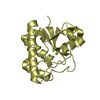
| ||||||||
| Unit cell |
| ||||||||
| Details | Biological assembly of this protein is monomer. |
-Components
| #1: Protein | Mass: 18467.926 Da / Num. of mol.: 4 Source method: isolated from a genetically manipulated source Source: (gene. exp.) Thermus thermophilus (bacteria) / Strain: HB8 / Plasmid: pET11a / Species (production host): Escherichia coli / Production host: #2: Chemical | ChemComp-MG /   Mass: 24.305 Da / Num. of mol.: 4 / Source method: obtained synthetically / Formula: Mg Mass: 24.305 Da / Num. of mol.: 4 / Source method: obtained synthetically / Formula: Mg#3: Water | ChemComp-HOH / |  Mass: 18.015 Da / Num. of mol.: 398 / Source method: isolated from a natural source / Formula: H2O Mass: 18.015 Da / Num. of mol.: 398 / Source method: isolated from a natural source / Formula: H2O |
|---|
-Experimental details
-Experiment
| Experiment | Method: X-RAY DIFFRACTION / Number of used crystals: 1 |
|---|
- Sample preparation
| Crystal | Density Matthews: 2.1 Å3/Da / Density % sol: 41.9 % |
|---|---|
| Crystal grow | Temperature: 295 K / Method: microbatch / pH: 6.5 Details: Magnesium acetate tetrahydrate, sodium cacodylate, PEG 8K, pH 6.5, microbatch, temperature 295K |
-Data collection
| Diffraction | Mean temperature: 100 K |
|---|---|
| Diffraction source | Source: SYNCHROTRON / Site: SPring-8  / Beamline: BL26B1 / Wavelength: 1 Å / Beamline: BL26B1 / Wavelength: 1 Å |
| Detector | Type: RIGAKU / Detector: IMAGE PLATE / Date: May 15, 2005 |
| Radiation | Monochromator: Graphite / Protocol: SINGLE WAVELENGTH / Monochromatic (M) / Laue (L): M / Scattering type: x-ray |
| Radiation wavelength | Wavelength: 1 Å / Relative weight: 1 |
| Reflection | Resolution: 1.9→50 Å / Num. all: 48911 / Num. obs: 48903 / % possible obs: 99.9 % / Observed criterion σ(F): 0 / Observed criterion σ(I): 0 / Redundancy: 3.4 % / Biso Wilson estimate: 18.9 Å2 / Rmerge(I) obs: 0.075 / Net I/σ(I): 7.3 |
| Reflection shell | Resolution: 1.9→1.97 Å / % possible all: 99.9 |
- Processing
| Software |
| |||||||||||||||||||||||||
|---|---|---|---|---|---|---|---|---|---|---|---|---|---|---|---|---|---|---|---|---|---|---|---|---|---|---|
| Refinement | Method to determine structure: MOLECULAR REPLACEMENT Starting model: 1DG9 Resolution: 1.9→38.04 Å / Rfactor Rfree error: 0.005 / Data cutoff high absF: 1585673.13 / Data cutoff low absF: 0 / Isotropic thermal model: RESTRAINED / Cross valid method: THROUGHOUT / σ(F): 0 / Stereochemistry target values: Engh & Huber
| |||||||||||||||||||||||||
| Solvent computation | Solvent model: FLAT MODEL / Bsol: 61.0453 Å2 / ksol: 0.393821 e/Å3 | |||||||||||||||||||||||||
| Displacement parameters | Biso mean: 32.2 Å2
| |||||||||||||||||||||||||
| Refine analyze |
| |||||||||||||||||||||||||
| Refinement step | Cycle: LAST / Resolution: 1.9→38.04 Å
| |||||||||||||||||||||||||
| Refine LS restraints |
| |||||||||||||||||||||||||
| LS refinement shell | Resolution: 1.9→2.02 Å / Rfactor Rfree error: 0.015 / Total num. of bins used: 6
| |||||||||||||||||||||||||
| Xplor file |
|
