 Movie
Movie Controller

[English] 日本語
 Yorodumi
- PDB-2cuy: Crystal structure of malonyl CoA-acyl carrier protein transacylas... -
+ Open data

- Basic information
| Entry | Database: PDB / ID: 2cuy | ||||||
|---|---|---|---|---|---|---|---|
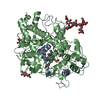
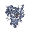
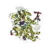
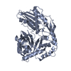
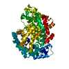
| Title | Crystal structure of malonyl CoA-acyl carrier protein transacylase from Thermus thermophilus HB8 | ||||||
 Components Components | Malonyl CoA-[acyl carrier protein] transacylase | ||||||
 Keywords Keywords | TRANSFERASE / Structural Genomics / NPPSFA / National Project on Protein Structural and Functional Analyses / RIKEN Structural Genomics/Proteomics Initiative / RSGI | ||||||
| Function / homology |  Function and homology information Function and homology information[acyl-carrier-protein] S-malonyltransferase / [acyl-carrier-protein] S-malonyltransferase activity / fatty acid biosynthetic process / cytosol Similarity search - Function | ||||||
| Biological species |   Thermus thermophilus (bacteria) Thermus thermophilus (bacteria) | ||||||
| Method |  X-RAY DIFFRACTION / SYNCHROTRON / MOLECULAR REPLACEMENT / Resolution: 2.1 Å X-RAY DIFFRACTION / SYNCHROTRON / MOLECULAR REPLACEMENT / Resolution: 2.1 Å | ||||||
 Authors Authors | Misaki, S. / Suzuki, K. / Kunishima, N. / Sugawara, M. / Kuroishi, C. / RIKEN Structural Genomics/Proteomics Initiative (RSGI) | ||||||
 Citation Citation | Journal: TO BE PUBLISHED Title: Crystal structure of malonyl CoA-acyl carrier protein transacylase from Thermus thermophilus HB8 Authors: Misaki, S. / Suzuki, K. / Kunishima, N. / Sugawara, M. / Kuroishi, C. | ||||||
| History |
|
- Structure visualization
| Structure viewer | Molecule: MolmilJmol/JSmol |
|---|
- Downloads & links
-Download
| PDBx/mmCIF format | 2cuy.cif.gz | 140.5 KB | Display | PDBx/mmCIF format |
|---|---|---|---|---|
| PDB format | pdb2cuy.ent.gz | 109.8 KB | Display | PDB format |
| PDBx/mmJSON format | 2cuy.json.gz | Tree view | PDBx/mmJSON format | |
| Others |  Other downloads Other downloads |
-Validation report
| Arichive directory | https://data.pdbj.org/pub/pdb/validation_reports/cu/2cuyftp://data.pdbj.org/pub/pdb/validation_reports/cu/2cuy | HTTPS FTP |
|---|
-Related structure data
| Related structure data |  1mlaS S: Starting model for refinement |
|---|---|
| Similar structure data | |
| Other databases |

-Links
 PDBj
PDBj
- Assembly
| Deposited unit | 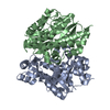
| ||||||||
|---|---|---|---|---|---|---|---|---|---|
| 1 |
| ||||||||
| Unit cell |
|
-Components
| #1: Protein | Mass: 33428.504 Da / Num. of mol.: 2 Source method: isolated from a genetically manipulated source Source: (gene. exp.) Thermus thermophilus (bacteria) / Strain: HB8 / Production host: References: UniProt: Q5SL77, [acyl-carrier-protein] S-malonyltransferase #2: Water | ChemComp-HOH / |  Mass: 18.015 Da / Num. of mol.: 672 / Source method: isolated from a natural source / Formula: H2O Mass: 18.015 Da / Num. of mol.: 672 / Source method: isolated from a natural source / Formula: H2O |
|---|
-Experimental details
-Experiment
| Experiment | Method: X-RAY DIFFRACTION / Number of used crystals: 1 |
|---|
- Sample preparation
| Crystal | Density Matthews: 3.68 Å3/Da / Density % sol: 66.62 % |
|---|---|
| Crystal grow | Temperature: 291 K / Method: evaporation / pH: 7.4 Details: magnesium sulfate, HEPES, pH 7.4, EVAPORATION, temperature 291K |
-Data collection
| Diffraction | Mean temperature: 100 K |
|---|---|
| Diffraction source | Source: SYNCHROTRON / Site: SPring-8  / Beamline: BL32B2 / Wavelength: 1 / Beamline: BL32B2 / Wavelength: 1 |
| Detector | Type: RIGAKU / Detector: IMAGE PLATE / Details: mirror |
| Radiation | Protocol: SINGLE WAVELENGTH / Monochromatic (M) / Laue (L): M / Scattering type: x-ray |
| Radiation wavelength | Wavelength: 1 Å / Relative weight: 1 |
| Reflection | Resolution: 2→50 Å / Num. obs: 63134 / % possible obs: 100 % / Observed criterion σ(I): 1 / Redundancy: 14.29 % / Rmerge(I) obs: 0.082 / Net I/σ(I): 19.4 |
| Reflection shell | Resolution: 2→2.07 Å / Redundancy: 14.44 % / Rmerge(I) obs: 0.317 / Mean I/σ(I) obs: 6.1 / % possible all: 100 |
- Processing
| Software |
| ||||||||||||||||||||
|---|---|---|---|---|---|---|---|---|---|---|---|---|---|---|---|---|---|---|---|---|---|
| Refinement | Method to determine structure: MOLECULAR REPLACEMENT Starting model: 1MLA Resolution: 2.1→30 Å / σ(F): 0 / Stereochemistry target values: Engh & Huber
| ||||||||||||||||||||
| Refinement step | Cycle: LAST / Resolution: 2.1→30 Å
|
