 Movie
Movie Controller

[English] 日本語
 Yorodumi
- PDB-1d7w: CRYSTAL STRUCTURE OF HUMAN MYELOPEROXIDASE ISOFORM C COMPLEXED WI... -
+ Open data

- Basic information
| Entry | Database: PDB / ID: 1d7w | ||||||||||||
|---|---|---|---|---|---|---|---|---|---|---|---|---|---|
| Title | CRYSTAL STRUCTURE OF HUMAN MYELOPEROXIDASE ISOFORM C COMPLEXED WITH CYANIDE AND BROMIDE AT PH 4.0 | ||||||||||||
 Components Components | (MYELOPEROXIDASE) x 2 | ||||||||||||
 Keywords Keywords | OXIDOREDUCTASE / HEME-PROTEIN / PEROXIDASE / PEROXIDASE-CYANIDE-BROMIDE COMPLEX | ||||||||||||
| Function / homology |  Function and homology information Function and homology informationmyeloperoxidase / hypochlorous acid biosynthetic process / Events associated with phagocytolytic activity of PMN cells / phagocytic vesicle lumen / response to gold nanoparticle / response to yeast / respiratory burst involved in defense response / low-density lipoprotein particle remodeling / azurophil granule / response to food ...myeloperoxidase / hypochlorous acid biosynthetic process / Events associated with phagocytolytic activity of PMN cells / phagocytic vesicle lumen / response to gold nanoparticle / response to yeast / respiratory burst involved in defense response / low-density lipoprotein particle remodeling / azurophil granule / response to food / defense response to fungus / response to mechanical stimulus / removal of superoxide radicals / secretory granule / hydrogen peroxide catabolic process / peroxidase activity / defense response / azurophil granule lumen / heparin binding / response to oxidative stress / response to lipopolysaccharide / lysosome / defense response to bacterium / heme binding / chromatin binding / Neutrophil degranulation / negative regulation of apoptotic process / : / extracellular exosome / extracellular region / nucleoplasm / metal ion binding / nucleus Similarity search - Function | ||||||||||||
| Biological species |  Homo sapiens (human) Homo sapiens (human) | ||||||||||||
| Method |  X-RAY DIFFRACTION / Resolution: 1.9 Å X-RAY DIFFRACTION / Resolution: 1.9 Å | ||||||||||||
 Authors Authors | Fiedler, T.J. / Fenna, R.E. | ||||||||||||
 Citation Citation | Journal: Biochemistry / Year: 2001 Title: Human myeloperoxidase: structure of a cyanide complex and its interaction with bromide and thiocyanate substrates at 1.9 A resolution. Authors: Blair-Johnson, M. / Fiedler, T. / Fenna, R. #1: Journal: Arch.Biochem.Biophys. / Year: 1995Title: Structure of the Green Heme in Myeloperoxidase Authors: Fenna, R. / Zeng, J. / Davey, C. #2: Journal: Biochemistry / Year: 1996Title: 2.3 Angstrom Resolution X-Ray Crystal Structure of the Bisubstrate Analogue Inhibitor Salicylhydroxamic Acid Bound to Human Myeloperoxidase: A Model for a Prereaction Complex with Hydrogen Peroxide Authors: Davey, C.A. / Fenna, R.E. #3: Journal: J.Mol.Biol. / Year: 1992Title: X-Ray Crystal Structure of Canine Myeloperoxidase at 3 Angstrom Resolution Authors: Zeng, J. / Fenna, R.E. #4: Journal: Biochem.Biophys.Res.Commun. / Year: 1994Title: Site-Directed Mutagenesis of Human Myeloperoxidase: Further Identification of Residues Involved in Catalytic Activity and Heme Interaction Authors: Jacquet, A. / Garcia-Quintana, L. / Deleersnyder, V. / Fenna, R. / Bollen, A. / Moguilevsky, N. | ||||||||||||
| History |
|
- Structure visualization
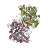
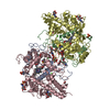
| Structure viewer | Molecule: MolmilJmol/JSmol |
|---|
- Downloads & links
-Download
| PDBx/mmCIF format | 1d7w.cif.gz | 268.6 KB | Display | PDBx/mmCIF format |
|---|---|---|---|---|
| PDB format | pdb1d7w.ent.gz | 211.3 KB | Display | PDB format |
| PDBx/mmJSON format | 1d7w.json.gz | Tree view | PDBx/mmJSON format | |
| Others |  Other downloads Other downloads |
-Validation report
| Arichive directory | https://data.pdbj.org/pub/pdb/validation_reports/d7/1d7wftp://data.pdbj.org/pub/pdb/validation_reports/d7/1d7w | HTTPS FTP |
|---|
-Related structure data


-Links
 PDBj
PDBj


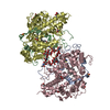
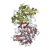
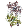
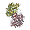
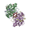
- Assembly
| Deposited unit | 
| ||||||||
|---|---|---|---|---|---|---|---|---|---|
| 1 |
| ||||||||
| 2 | 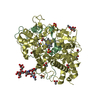
| ||||||||
| 3 | 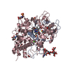
| ||||||||
| Unit cell |
| ||||||||
| Noncrystallographic symmetry (NCS) | NCS oper: (Code: given Matrix: (-0.6064, 0.68136, -0.4099), Vector: |
-Components
-Protein , 2 types, 4 molecules ABCD
| #1: Protein | Mass: 11903.343 Da / Num. of mol.: 2 / Fragment: LIGHT CHAIN / Source method: isolated from a natural source / Source: (natural) Homo sapiens (human) / Cell: NEUTROPHIL / Tissue: BLOOD / References: UniProt: P05164, peroxidase#2: Protein | Mass: 53234.191 Da / Num. of mol.: 2 / Fragment: HEAVY CHAIN / Source method: isolated from a natural source / Source: (natural) Homo sapiens (human) / Cell: NEUTROPHIL / Tissue: BLOOD / References: UniProt: P05164, peroxidase |
|---|
-Sugars , 2 types, 6 molecules 
| #3: Polysaccharide | Source method: isolated from a genetically manipulated source #9: Sugar | ChemComp-NAG /  Type: D-saccharide, beta linking / Mass: 221.208 Da / Num. of mol.: 4 Type: D-saccharide, beta linking / Mass: 221.208 Da / Num. of mol.: 4Source method: isolated from a genetically manipulated source Formula: C8H15NO6 |
|---|
-Non-polymers , 7 types, 837 molecules 


| #4: Chemical | ChemComp-BR /  Mass: 79.904 Da / Num. of mol.: 8 / Source method: obtained synthetically / Formula: Br Mass: 79.904 Da / Num. of mol.: 8 / Source method: obtained synthetically / Formula: Br#5: Chemical | ChemComp-SO4 /  Mass: 96.063 Da / Num. of mol.: 5 / Source method: obtained synthetically / Formula: SO4 Mass: 96.063 Da / Num. of mol.: 5 / Source method: obtained synthetically / Formula: SO4#6: Chemical |  Mass: 26.017 Da / Num. of mol.: 2 / Source method: obtained synthetically / Formula: CN Mass: 26.017 Da / Num. of mol.: 2 / Source method: obtained synthetically / Formula: CN#7: Chemical | ChemComp-ACT /  Mass: 59.044 Da / Num. of mol.: 12 / Source method: obtained synthetically / Formula: C2H3O2 Mass: 59.044 Da / Num. of mol.: 12 / Source method: obtained synthetically / Formula: C2H3O2#8: Chemical |  Mass: 616.487 Da / Num. of mol.: 2 / Source method: obtained synthetically / Formula: C34H32FeN4O4 Mass: 616.487 Da / Num. of mol.: 2 / Source method: obtained synthetically / Formula: C34H32FeN4O4#10: Chemical |  Mass: 40.078 Da / Num. of mol.: 2 / Source method: obtained synthetically / Formula: Ca Mass: 40.078 Da / Num. of mol.: 2 / Source method: obtained synthetically / Formula: Ca#11: Water | ChemComp-HOH / | Mass: 18.015 Da / Num. of mol.: 806 / Source method: isolated from a natural source / Formula: H2O |
|---|
-Details
| Has protein modification | Y |
|---|
-Experimental details
-Experiment
| Experiment | Method: X-RAY DIFFRACTION / Number of used crystals: 1 |
|---|
- Sample preparation
| Crystal | Density Matthews: 2.47 Å3/Da / Density % sol: 50.28 % | |||||||||||||||||||||||||||||||||||||||||||||||||
|---|---|---|---|---|---|---|---|---|---|---|---|---|---|---|---|---|---|---|---|---|---|---|---|---|---|---|---|---|---|---|---|---|---|---|---|---|---|---|---|---|---|---|---|---|---|---|---|---|---|---|
| Crystal grow | Temperature: 289 K / Method: vapor diffusion, hanging drop / pH: 4 Details: polyethylene glycol 8000, ammonium sulfate, sodium acetate, calcium acetate, sodium cyanide, sodium bromide, pH 4.0, VAPOR DIFFUSION, HANGING DROP, temperature 289K | |||||||||||||||||||||||||||||||||||||||||||||||||
| Crystal grow | *PLUS pH: 5 / Details: Davey, C.A., (1996) Biochemistry, 35, 10967. | |||||||||||||||||||||||||||||||||||||||||||||||||
| Components of the solutions | *PLUS
|
-Data collection
| Diffraction | Mean temperature: 84 K |
|---|---|
| Diffraction source | Source: ROTATING ANODE / Type: RIGAKU RU300 / Wavelength: 1.5418 |
| Detector | Type: MARRESEARCH / Detector: IMAGE PLATE / Date: Jun 21, 1999 / Details: LONG FOCUSING MIRRORS, ADSC |
| Radiation | Protocol: SINGLE WAVELENGTH / Monochromatic (M) / Laue (L): M / Scattering type: x-ray |
| Radiation wavelength | Wavelength: 1.5418 Å / Relative weight: 1 |
| Reflection | Resolution: 1.9→50 Å / Num. obs: 92679 / % possible obs: 92.3 % / Observed criterion σ(I): 0 / Redundancy: 3.71 % / Biso Wilson estimate: 15.25 Å2 / Rmerge(I) obs: 0.061 / Net I/σ(I): 7.89 |
| Reflection shell | Resolution: 1.9→1.97 Å / Redundancy: 3.89 % / Rmerge(I) obs: 0.191 / Mean I/σ(I) obs: 7.89 / % possible all: 89.8 |
| Reflection | *PLUS Num. measured all: 343921 |
| Reflection shell | *PLUS % possible obs: 89.8 % |
- Processing
| Software |
| ||||||||||||||||||||||||||||||||||||||||||||||||||||||||||||
|---|---|---|---|---|---|---|---|---|---|---|---|---|---|---|---|---|---|---|---|---|---|---|---|---|---|---|---|---|---|---|---|---|---|---|---|---|---|---|---|---|---|---|---|---|---|---|---|---|---|---|---|---|---|---|---|---|---|---|---|---|---|
| Refinement | Resolution: 1.9→30 Å / σ(F): 2
| ||||||||||||||||||||||||||||||||||||||||||||||||||||||||||||
| Refine analyze |
| ||||||||||||||||||||||||||||||||||||||||||||||||||||||||||||
| Refinement step | Cycle: LAST / Resolution: 1.9→30 Å
| ||||||||||||||||||||||||||||||||||||||||||||||||||||||||||||
| Refine LS restraints |
| ||||||||||||||||||||||||||||||||||||||||||||||||||||||||||||
| LS refinement shell | Resolution: 1.9→1.97 Å / Total num. of bins used: 10
| ||||||||||||||||||||||||||||||||||||||||||||||||||||||||||||
| Software | *PLUS Name: X-PLOR / Version: 3.851 / Classification: refinement | ||||||||||||||||||||||||||||||||||||||||||||||||||||||||||||
| Refinement | *PLUS % reflection Rfree: 5 % / Rfactor Rfree: 0.27 | ||||||||||||||||||||||||||||||||||||||||||||||||||||||||||||
| Solvent computation | *PLUS | ||||||||||||||||||||||||||||||||||||||||||||||||||||||||||||
| Displacement parameters | *PLUS | ||||||||||||||||||||||||||||||||||||||||||||||||||||||||||||
| Refine LS restraints | *PLUS
| ||||||||||||||||||||||||||||||||||||||||||||||||||||||||||||
| LS refinement shell | *PLUS Rfactor Rwork: 0.271 / Rfactor obs: 0.271 |
