 Movie
Movie Controller

[English] 日本語
 Yorodumi
- PDB-2cun: Crystal structure of Phosphoglycerate Kinase from Pyrococcus hori... -
+ Open data

- Basic information
| Entry | Database: PDB / ID: 2cun | ||||||
|---|---|---|---|---|---|---|---|
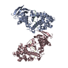
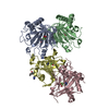
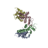
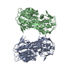
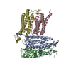
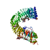
| Title | Crystal structure of Phosphoglycerate Kinase from Pyrococcus horikoshii OT3 | ||||||
 Components Components | Phosphoglycerate kinase | ||||||
 Keywords Keywords | TRANSFERASE / Phosphoglycerate kinase / Structural Genomics / TANPAKU 3000 / RIKEN Structural Genomics/Proteomics Initiative / RSGI / NPPSFA / National Project on Protein Structural and Functional Analyses | ||||||
| Function / homology |  Function and homology information Function and homology informationphosphoglycerate kinase / phosphoglycerate kinase activity / gluconeogenesis / glycolytic process / ADP binding / ATP binding / cytosol Similarity search - Function | ||||||
| Biological species |   Pyrococcus horikoshii (archaea) Pyrococcus horikoshii (archaea) | ||||||
| Method |  X-RAY DIFFRACTION / SYNCHROTRON / MOLECULAR REPLACEMENT / Resolution: 2.1 Å X-RAY DIFFRACTION / SYNCHROTRON / MOLECULAR REPLACEMENT / Resolution: 2.1 Å | ||||||
 Authors Authors | Mizutani, H. / Kunishima, N. / RIKEN Structural Genomics/Proteomics Initiative (RSGI) | ||||||
 Citation Citation | Journal: To be Published Title: Crystal structure of Phosphoglycerate Kinase from Pyrococcus horikoshii OT3 Authors: Mizutani, H. / Kunishima, N. | ||||||
| History |
|
- Structure visualization
| Structure viewer | Molecule: MolmilJmol/JSmol |
|---|
- Downloads & links
-Download
| PDBx/mmCIF format | 2cun.cif.gz | 177.9 KB | Display | PDBx/mmCIF format |
|---|---|---|---|---|
| PDB format | pdb2cun.ent.gz | 141 KB | Display | PDB format |
| PDBx/mmJSON format | 2cun.json.gz | Tree view | PDBx/mmJSON format | |
| Others |  Other downloads Other downloads |
-Validation report
| Arichive directory | https://data.pdbj.org/pub/pdb/validation_reports/cu/2cunftp://data.pdbj.org/pub/pdb/validation_reports/cu/2cun | HTTPS FTP |
|---|
-Related structure data
| Related structure data | 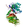 1vpeS S: Starting model for refinement |
|---|---|
| Similar structure data | |
| Other databases |


-Links
 PDBj
PDBj
- Assembly
| Deposited unit | 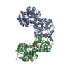
| ||||||||
|---|---|---|---|---|---|---|---|---|---|
| 1 |
| ||||||||
| Unit cell |
| ||||||||
| Details | The biological assembly is a dimer, chain A & B in asymmetric unit. |
-Components
-Protein , 1 types, 2 molecules AB
| #1: Protein | Mass: 46470.680 Da / Num. of mol.: 2 Source method: isolated from a genetically manipulated source Source: (gene. exp.) Pyrococcus horikoshii (archaea) / Strain: OT3 / Plasmid: pET11a / Production host:  |
|---|
-Non-polymers , 5 types, 380 molecules 


| #2: Chemical |  Mass: 35.453 Da / Num. of mol.: 2 / Source method: obtained synthetically / Formula: Cl Mass: 35.453 Da / Num. of mol.: 2 / Source method: obtained synthetically / Formula: Cl#3: Chemical |  Mass: 186.057 Da / Num. of mol.: 2 / Source method: obtained synthetically / Formula: C3H7O7P Mass: 186.057 Da / Num. of mol.: 2 / Source method: obtained synthetically / Formula: C3H7O7P#4: Chemical |  Mass: 92.094 Da / Num. of mol.: 2 / Source method: obtained synthetically / Formula: C3H8O3 Mass: 92.094 Da / Num. of mol.: 2 / Source method: obtained synthetically / Formula: C3H8O3#5: Chemical |  Mass: 118.174 Da / Num. of mol.: 3 / Source method: obtained synthetically / Formula: C6H14O2 / Comment: precipitant*YM Mass: 118.174 Da / Num. of mol.: 3 / Source method: obtained synthetically / Formula: C6H14O2 / Comment: precipitant*YM#6: Water | ChemComp-HOH / | Mass: 18.015 Da / Num. of mol.: 371 / Source method: isolated from a natural source / Formula: H2O |
|---|
-Experimental details
-Experiment
| Experiment | Method: X-RAY DIFFRACTION / Number of used crystals: 1 |
|---|
- Sample preparation
| Crystal | Density Matthews: 2.74 Å3/Da / Density % sol: 55.11 % |
|---|---|
| Crystal grow | Temperature: 295 K / Method: microbatch / pH: 7.5 Details: PEG 6000, MPD, HEPES, pH 7.5, microbatch, temperature 295K |
-Data collection
| Diffraction | Mean temperature: 100 K |
|---|---|
| Diffraction source | Source: SYNCHROTRON / Site: SPring-8  / Beamline: BL26B1 / Wavelength: 1 Å / Beamline: BL26B1 / Wavelength: 1 Å |
| Detector | Type: RIGAKU JUPITER 210 / Detector: CCD / Date: Oct 1, 2004 / Details: mirrors |
| Radiation | Monochromator: Bending magnet / Protocol: SINGLE WAVELENGTH / Monochromatic (M) / Laue (L): M / Scattering type: x-ray |
| Radiation wavelength | Wavelength: 1 Å / Relative weight: 1 |
| Reflection | Resolution: 2.1→30 Å / Num. all: 58103 / Num. obs: 58103 / % possible obs: 100 % / Observed criterion σ(F): 0 / Observed criterion σ(I): 0 / Redundancy: 4.6 % / Biso Wilson estimate: 25.2 Å2 / Rmerge(I) obs: 0.068 / Rsym value: 0.054 / Net I/σ(I): 12.1 |
| Reflection shell | Resolution: 2.1→2.18 Å / Redundancy: 4.5 % / Rmerge(I) obs: 0.3 / Mean I/σ(I) obs: 3.27 / Num. unique all: 5810 / Rsym value: 0.256 / % possible all: 99.9 |
- Processing
| Software |
| ||||||||||||||||||||||||||||||||||||||||||||||||||||||||||||||||||||||||||||||||
|---|---|---|---|---|---|---|---|---|---|---|---|---|---|---|---|---|---|---|---|---|---|---|---|---|---|---|---|---|---|---|---|---|---|---|---|---|---|---|---|---|---|---|---|---|---|---|---|---|---|---|---|---|---|---|---|---|---|---|---|---|---|---|---|---|---|---|---|---|---|---|---|---|---|---|---|---|---|---|---|---|---|
| Refinement | Method to determine structure: MOLECULAR REPLACEMENT Starting model: PDB entry 1VPE Resolution: 2.1→30 Å / Rfactor Rfree error: 0.005 / Data cutoff high absF: 1594550.5 / Data cutoff low absF: 0 / Isotropic thermal model: RESTRAINED / Cross valid method: THROUGHOUT / σ(F): 0 / Stereochemistry target values: Engh & Huber
| ||||||||||||||||||||||||||||||||||||||||||||||||||||||||||||||||||||||||||||||||
| Solvent computation | Solvent model: FLAT MODEL / Bsol: 54.0678 Å2 / ksol: 0.338601 e/Å3 | ||||||||||||||||||||||||||||||||||||||||||||||||||||||||||||||||||||||||||||||||
| Displacement parameters | Biso mean: 44.2 Å2
| ||||||||||||||||||||||||||||||||||||||||||||||||||||||||||||||||||||||||||||||||
| Refine analyze |
| ||||||||||||||||||||||||||||||||||||||||||||||||||||||||||||||||||||||||||||||||
| Refinement step | Cycle: LAST / Resolution: 2.1→30 Å
| ||||||||||||||||||||||||||||||||||||||||||||||||||||||||||||||||||||||||||||||||
| Refine LS restraints |
| ||||||||||||||||||||||||||||||||||||||||||||||||||||||||||||||||||||||||||||||||
| LS refinement shell | Resolution: 2.1→2.23 Å / Rfactor Rfree error: 0.014 / Total num. of bins used: 6
| ||||||||||||||||||||||||||||||||||||||||||||||||||||||||||||||||||||||||||||||||
| Xplor file |
|
