 Movie
Movie Controller

[English] 日本語
 Yorodumi
- PDB-2ci5: Crystal structure of Dimethylarginine Dimethylaminohydrolase I in... -
+ Open data

- Basic information
| Entry | Database: PDB / ID: 2ci5 | |||||||||
|---|---|---|---|---|---|---|---|---|---|---|
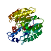
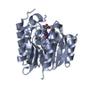
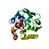
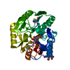
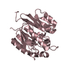
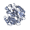
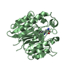
| Title | Crystal structure of Dimethylarginine Dimethylaminohydrolase I in complex with L-homocysteine | |||||||||
 Components Components | NG, NG-DIMETHYLARGININE DIMETHYLAMINOHYDROLASE 1 | |||||||||
 Keywords Keywords | HYDROLASE / NOS REGULATION / S-NITROSYLATION / ZINC / NO / MMA / ADMA / ACETYLATION / METAL-BINDING | |||||||||
| Function / homology |  Function and homology information Function and homology informationprotein nitrosylation / eNOS activation / dimethylargininase / dimethylargininase activity / : / arginine metabolic process / amino acid binding / nitric oxide biosynthetic process / positive regulation of nitric oxide biosynthetic process / zinc ion binding Similarity search - Function | |||||||||
| Biological species |  | |||||||||
| Method |  X-RAY DIFFRACTION / SYNCHROTRON / MOLECULAR REPLACEMENT / Resolution: 1.79 Å X-RAY DIFFRACTION / SYNCHROTRON / MOLECULAR REPLACEMENT / Resolution: 1.79 Å | |||||||||
 Authors Authors | Frey, D. / Braun, O. / Briand, C. / Vasak, M. / Grutter, M.G. | |||||||||
 Citation Citation | Journal: Structure / Year: 2006 Title: Structure of the Mammalian Nos Regulator Dimethylarginine Dimethylaminohydrolase: A Basis for the Design of Specific Inbitors Authors: Frey, D. / Braun, O. / Briand, C. / Vasak, M. / Grutter, M.G. | |||||||||
| History |
|
- Structure visualization
| Structure viewer | Molecule: MolmilJmol/JSmol |
|---|
- Downloads & links
-Download
| PDBx/mmCIF format | 2ci5.cif.gz | 129.5 KB | Display | PDBx/mmCIF format |
|---|---|---|---|---|
| PDB format | pdb2ci5.ent.gz | 101.2 KB | Display | PDB format |
| PDBx/mmJSON format | 2ci5.json.gz | Tree view | PDBx/mmJSON format | |
| Others |  Other downloads Other downloads |
-Validation report
| Arichive directory | https://data.pdbj.org/pub/pdb/validation_reports/ci/2ci5ftp://data.pdbj.org/pub/pdb/validation_reports/ci/2ci5 | HTTPS FTP |
|---|
-Related structure data
| Related structure data | 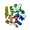 2c6zC  2ci1C 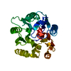 2ci3C  2ci4C  2ci6C 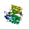 2ci7C 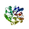 1h70S C: citing same article ( S: Starting model for refinement |
|---|---|
| Similar structure data |


-Links
 PDBj
PDBj- Assembly
| Deposited unit | 
| ||||||||
|---|---|---|---|---|---|---|---|---|---|
| 1 | 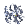
| ||||||||
| 2 | 
| ||||||||
| Unit cell |
| ||||||||
| Noncrystallographic symmetry (NCS) | NCS oper: (Code: given Matrix: (0.99999, 0.00489, -0.0023), Vector: |
-Components
| #1: Protein | Mass: 31198.672 Da / Num. of mol.: 2 / Source method: isolated from a natural source / Source: (natural) #2: Chemical |   Type: L-peptide linking / Mass: 135.185 Da / Num. of mol.: 2 / Source method: obtained synthetically / Formula: C4H9NO2S Type: L-peptide linking / Mass: 135.185 Da / Num. of mol.: 2 / Source method: obtained synthetically / Formula: C4H9NO2S#3: Chemical | ChemComp-CIT / |   Mass: 192.124 Da / Num. of mol.: 1 / Source method: obtained synthetically / Formula: C6H8O7 Mass: 192.124 Da / Num. of mol.: 1 / Source method: obtained synthetically / Formula: C6H8O7#4: Water | ChemComp-HOH / |  Mass: 18.015 Da / Num. of mol.: 647 / Source method: isolated from a natural source / Formula: H2O Mass: 18.015 Da / Num. of mol.: 647 / Source method: isolated from a natural source / Formula: H2O |
|---|
-Experimental details
-Experiment
| Experiment | Method: X-RAY DIFFRACTION / Number of used crystals: 1 |
|---|
- Sample preparation
| Crystal | Density Matthews: 2.16 Å3/Da / Density % sol: 43 % |
|---|---|
| Crystal grow | pH: 5 Details: 100 MM CITRIC ACID/NAOH, 20-40% PEG 8000, 2 MM TCEP, PH 5.0 |
-Data collection
| Diffraction | Mean temperature: 100 K |
|---|---|
| Diffraction source | Source: SYNCHROTRON / Site: SLS  / Beamline: X06SA / Wavelength: 0.9793 / Beamline: X06SA / Wavelength: 0.9793 |
| Detector | Type: MARRESEARCH / Detector: CCD / Date: Nov 11, 2003 / Details: DYNAMICALLY BENDABLE MIRROR |
| Radiation | Monochromator: SI(111) / Protocol: SINGLE WAVELENGTH / Monochromatic (M) / Laue (L): M / Scattering type: x-ray |
| Radiation wavelength | Wavelength: 0.9793 Å / Relative weight: 1 |
| Reflection | Resolution: 1.8→30 Å / Num. obs: 48415 / % possible obs: 98.1 % / Observed criterion σ(I): 2 / Redundancy: 3.25 % / Biso Wilson estimate: 11 Å2 / Rmerge(I) obs: 0.13 / Net I/σ(I): 9.5 |
| Reflection shell | Resolution: 1.8→1.86 Å / Rmerge(I) obs: 0.38 / Mean I/σ(I) obs: 2.2 / % possible all: 90.7 |
- Processing
| Software |
| ||||||||||||||||||||||||||||||||||||||||||||||||||||||||||||||||||||||||||||||||
|---|---|---|---|---|---|---|---|---|---|---|---|---|---|---|---|---|---|---|---|---|---|---|---|---|---|---|---|---|---|---|---|---|---|---|---|---|---|---|---|---|---|---|---|---|---|---|---|---|---|---|---|---|---|---|---|---|---|---|---|---|---|---|---|---|---|---|---|---|---|---|---|---|---|---|---|---|---|---|---|---|---|
| Refinement | Method to determine structure: MOLECULAR REPLACEMENT Starting model: PDB ENTRY 1H70 Resolution: 1.79→29.2 Å / Rfactor Rfree error: 0.004 / Data cutoff high absF: 1453876.18 / Isotropic thermal model: RESTRAINED / Cross valid method: THROUGHOUT / σ(F): 0 / Stereochemistry target values: MAXIMUM LIKELIHOOD
| ||||||||||||||||||||||||||||||||||||||||||||||||||||||||||||||||||||||||||||||||
| Solvent computation | Solvent model: FLAT MODEL / Bsol: 34.7133 Å2 / ksol: 0.348887 e/Å3 | ||||||||||||||||||||||||||||||||||||||||||||||||||||||||||||||||||||||||||||||||
| Displacement parameters | Biso mean: 15 Å2
| ||||||||||||||||||||||||||||||||||||||||||||||||||||||||||||||||||||||||||||||||
| Refine analyze |
| ||||||||||||||||||||||||||||||||||||||||||||||||||||||||||||||||||||||||||||||||
| Refinement step | Cycle: LAST / Resolution: 1.79→29.2 Å
| ||||||||||||||||||||||||||||||||||||||||||||||||||||||||||||||||||||||||||||||||
| Refine LS restraints |
| ||||||||||||||||||||||||||||||||||||||||||||||||||||||||||||||||||||||||||||||||
| LS refinement shell | Resolution: 1.7→1.81 Å / Rfactor Rfree error: 0.085 / Total num. of bins used: 6
| ||||||||||||||||||||||||||||||||||||||||||||||||||||||||||||||||||||||||||||||||
| Xplor file |
|
