 Movie
Movie Controller

[English] 日本語
 Yorodumi
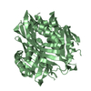
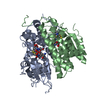
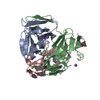
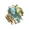
- PDB-2chc: Structure of Rv3472(D26N), a function unknown protein from Mycoba... -
+ Open data

- Basic information
| Entry | Database: PDB / ID: 2chc | ||||||
|---|---|---|---|---|---|---|---|
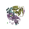
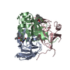
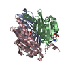
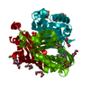
| Title | Structure of Rv3472(D26N), a function unknown protein from Mycobacterium tuberculosis | ||||||
 Components Components | PROTEIN RV3472 | ||||||
 Keywords Keywords | HYPOTHETICAL PROTEIN / RV3472 / MYCOBACTERIUM / TUBERCULOSIS | ||||||
| Function / homology | SnoaL-like domain / SnoaL-like domain / Nuclear Transport Factor 2; Chain: A, - #50 / NTF2-like domain superfamily / Nuclear Transport Factor 2; Chain: A, / Roll / Alpha Beta / SnoaL-like domain-containing protein Function and homology information Function and homology information | ||||||
| Biological species |   MYCOBACTERIUM TUBERCULOSIS (bacteria) MYCOBACTERIUM TUBERCULOSIS (bacteria) | ||||||
| Method |  X-RAY DIFFRACTION / SYNCHROTRON / SAD / Resolution: 1.69 Å X-RAY DIFFRACTION / SYNCHROTRON / SAD / Resolution: 1.69 Å | ||||||
 Authors Authors | Ma, Q. / Wilmanns, M. | ||||||
 Citation Citation | Journal: To be Published Title: Structure of Rv3472(D26N), a Function Unknown Protein from Mycobacterium Tuberculosis Authors: Ma, Q. / Wilmanns, M. | ||||||
| History |
|
- Structure visualization
| Structure viewer | Molecule: MolmilJmol/JSmol |
|---|
- Downloads & links
-Download
| PDBx/mmCIF format | 2chc.cif.gz | 109.1 KB | Display | PDBx/mmCIF format |
|---|---|---|---|---|
| PDB format | pdb2chc.ent.gz | 86.4 KB | Display | PDB format |
| PDBx/mmJSON format | 2chc.json.gz | Tree view | PDBx/mmJSON format | |
| Others |  Other downloads Other downloads |
-Validation report
| Summary document | 2chc_validation.pdf.gz | 448.4 KB | Display | wwPDB validaton report |
|---|---|---|---|---|
| Full document | 2chc_full_validation.pdf.gz | 456.7 KB | Display | |
| Data in XML | 2chc_validation.xml.gz | 22.9 KB | Display | |
| Data in CIF | 2chc_validation.cif.gz | 33.4 KB | Display | |
| Arichive directory | https://data.pdbj.org/pub/pdb/validation_reports/ch/2chcftp://data.pdbj.org/pub/pdb/validation_reports/ch/2chc | HTTPS FTP |
-Related structure data
| Similar structure data |
|---|
-Links
 PDBj
PDBj
- Assembly
| Deposited unit | 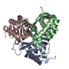
| ||||||||
|---|---|---|---|---|---|---|---|---|---|
| 1 |
| ||||||||
| Unit cell |
|
-Components
| #1: Protein | Mass: 18617.898 Da / Num. of mol.: 3 / Mutation: YES Source method: isolated from a genetically manipulated source Source: (gene. exp.) MYCOBACTERIUM TUBERCULOSIS (bacteria) / Strain: H37RV / Plasmid: PETM11 / Production host: #2: Water | ChemComp-HOH / |  Mass: 18.015 Da / Num. of mol.: 327 / Source method: isolated from a natural source / Formula: H2O Mass: 18.015 Da / Num. of mol.: 327 / Source method: isolated from a natural source / Formula: H2OCompound details | ENGINEERED RESIDUE IN CHAIN A, ASP 26 TO ASN ENGINEERED RESIDUE IN CHAIN B, ASP 26 TO ASN ...ENGINEERED | Sequence details | SOME RESIDUES ARE ALTERED DUE TO GENE CONSTRUCTION METHOD. ATOMS WITH VERY POOR ELECTRON DENSITY ...SOME RESIDUES ARE ALTERED DUE TO GENE CONSTRUCTI | |
|---|
-Experimental details
-Experiment
| Experiment | Method: X-RAY DIFFRACTION / Number of used crystals: 1 |
|---|
- Sample preparation
| Crystal | Density Matthews: 2.4 Å3/Da / Density % sol: 48 % |
|---|---|
| Crystal grow | Temperature: 293 K / pH: 7.4 Details: 0.1M HEPES PH7.4, 0.2M MGCL2, 25% PEG400, 20 DEGREES C, pH 7.40 |
-Data collection
| Diffraction | Mean temperature: 100 K |
|---|---|
| Diffraction source | Source: SYNCHROTRON / Site: EMBL/DESY, HAMBURG  / Beamline: BW7B / Wavelength: 0.8424 / Beamline: BW7B / Wavelength: 0.8424 |
| Detector | Type: MARRESEARCH / Detector: IMAGE PLATE / Date: May 1, 2005 / Details: MIRROR |
| Radiation | Monochromator: TRIANGULAR / Protocol: SINGLE WAVELENGTH / Monochromatic (M) / Laue (L): M / Scattering type: x-ray |
| Radiation wavelength | Wavelength: 0.8424 Å / Relative weight: 1 |
| Reflection | Resolution: 1.69→37.3 Å / Num. obs: 60209 / % possible obs: 99.6 % / Redundancy: 6.5 % / Biso Wilson estimate: 23.6 Å2 / Rmerge(I) obs: 0.04 / Net I/σ(I): 24.67 |
| Reflection shell | Resolution: 1.69→1.79 Å / Redundancy: 5.71 % / Rmerge(I) obs: 0.32 / Mean I/σ(I) obs: 5.23 / % possible all: 98.6 |
- Processing
| Software |
| ||||||||||||||||||||||||||||||||||||||||||||||||||||||||||||||||||||||||||||||||||||||||||||||||||||||||||||||||||||||||||||||||||||||||||||||||||||||||||||||||||||||||||||||||||||||
|---|---|---|---|---|---|---|---|---|---|---|---|---|---|---|---|---|---|---|---|---|---|---|---|---|---|---|---|---|---|---|---|---|---|---|---|---|---|---|---|---|---|---|---|---|---|---|---|---|---|---|---|---|---|---|---|---|---|---|---|---|---|---|---|---|---|---|---|---|---|---|---|---|---|---|---|---|---|---|---|---|---|---|---|---|---|---|---|---|---|---|---|---|---|---|---|---|---|---|---|---|---|---|---|---|---|---|---|---|---|---|---|---|---|---|---|---|---|---|---|---|---|---|---|---|---|---|---|---|---|---|---|---|---|---|---|---|---|---|---|---|---|---|---|---|---|---|---|---|---|---|---|---|---|---|---|---|---|---|---|---|---|---|---|---|---|---|---|---|---|---|---|---|---|---|---|---|---|---|---|---|---|---|---|
| Refinement | Method to determine structure: SAD / Resolution: 1.69→37.32 Å / Cor.coef. Fo:Fc: 0.966 / Cor.coef. Fo:Fc free: 0.954 / SU B: 3.356 / SU ML: 0.056 / TLS residual ADP flag: LIKELY RESIDUAL / Cross valid method: THROUGHOUT / ESU R: 0.088 / ESU R Free: 0.091 / Stereochemistry target values: MAXIMUM LIKELIHOOD / Details: HYDROGENS HAVE BEEN ADDED IN THE RIDING POSITIONS.
| ||||||||||||||||||||||||||||||||||||||||||||||||||||||||||||||||||||||||||||||||||||||||||||||||||||||||||||||||||||||||||||||||||||||||||||||||||||||||||||||||||||||||||||||||||||||
| Solvent computation | Ion probe radii: 0.8 Å / Shrinkage radii: 0.8 Å / VDW probe radii: 1.2 Å / Solvent model: BABINET MODEL WITH MASK | ||||||||||||||||||||||||||||||||||||||||||||||||||||||||||||||||||||||||||||||||||||||||||||||||||||||||||||||||||||||||||||||||||||||||||||||||||||||||||||||||||||||||||||||||||||||
| Displacement parameters | Biso mean: 22.68 Å2
| ||||||||||||||||||||||||||||||||||||||||||||||||||||||||||||||||||||||||||||||||||||||||||||||||||||||||||||||||||||||||||||||||||||||||||||||||||||||||||||||||||||||||||||||||||||||
| Refinement step | Cycle: LAST / Resolution: 1.69→37.32 Å
| ||||||||||||||||||||||||||||||||||||||||||||||||||||||||||||||||||||||||||||||||||||||||||||||||||||||||||||||||||||||||||||||||||||||||||||||||||||||||||||||||||||||||||||||||||||||
| Refine LS restraints |
|