 Movie
Movie Controller


+ Open data

- Basic information
| Entry | Database: PDB / ID: 2cf2 | ||||||
|---|---|---|---|---|---|---|---|
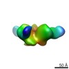
| Title | Architecture of mammalian fatty acid synthase | ||||||
 Components Components |
| ||||||
 Keywords Keywords | TRANSFERASE / FATTY ACID METABOLISM / FATTY ACID BIOSYNTHESIS / MULTIENZYME | ||||||
| Function / homology |  Function and homology information Function and homology informationmonounsaturated fatty acid biosynthetic process / beta-ketoacyl-[acyl-carrier-protein] synthase I / 3-oxoacyl-[acyl-carrier-protein] synthase activity / fatty acid biosynthetic process / cytosol Similarity search - Function | ||||||
| Biological species |  | ||||||
| Method |  X-RAY DIFFRACTION / SYNCHROTRON / MIRAS / Resolution: 4.3 Å X-RAY DIFFRACTION / SYNCHROTRON / MIRAS / Resolution: 4.3 Å | ||||||
| Model type details | CA ATOMS ONLY, CHAIN A, J, B, K, C, L, D, M, E, N | ||||||
 Authors Authors | Maier, T. / Jenni, S. / Ban, N. | ||||||
 Citation Citation | Journal: Science / Year: 2006 Title: Architecture of Mammalian Fatty Acid Synthase at 4.5 A Resolution. Authors: Maier, T. / Jenni, S. / Ban, N. | ||||||
| History |
|
- Structure visualization
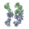
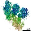
| Structure viewer | Molecule: MolmilJmol/JSmol |
|---|
- Downloads & links
-Download
| PDBx/mmCIF format | 2cf2.cif.gz | 110.9 KB | Display | PDBx/mmCIF format |
|---|---|---|---|---|
| PDB format | pdb2cf2.ent.gz | 62.6 KB | Display | PDB format |
| PDBx/mmJSON format | 2cf2.json.gz | Tree view | PDBx/mmJSON format | |
| Others |  Other downloads Other downloads |
-Validation report
| Arichive directory | https://data.pdbj.org/pub/pdb/validation_reports/cf/2cf2ftp://data.pdbj.org/pub/pdb/validation_reports/cf/2cf2 | HTTPS FTP |
|---|
-Related structure data
| Related structure data | |
|---|---|
| Similar structure data |


-Links
 PDBj
PDBj

- Assembly
| Deposited unit | 
| ||||||||
|---|---|---|---|---|---|---|---|---|---|
| 1 |
| ||||||||
| Unit cell |
|
-Components
| #1: Protein | Mass: 42668.191 Da / Num. of mol.: 2 / Source method: isolated from a natural source / Source: (natural) References: UniProt: P0A953*PLUS, fatty-acid synthase system #2: Protein | Mass: 30981.406 Da / Num. of mol.: 2 / Source method: isolated from a natural source / Source: (natural) #3: Protein | Mass: 37701.637 Da / Num. of mol.: 2 / Source method: isolated from a natural source / Source: (natural) #4: Protein | Mass: 31513.039 Da / Num. of mol.: 2 / Source method: isolated from a natural source / Source: (natural) #5: Protein | Mass: 23702.061 Da / Num. of mol.: 2 / Source method: isolated from a natural source / Source: (natural) Sequence details | THIS PDB FILE CONTAINS INDIVIDUAL HOMOLOGOUS DOMAINS PLACED INTO AN EXPERIMENTALLY PHASED 4.5A ...THIS PDB FILE CONTAINS INDIVIDUAL | |
|---|
-Experimental details
-Experiment
| Experiment | Method: X-RAY DIFFRACTION / Number of used crystals: 1 |
|---|
- Sample preparation
| Crystal | Density Matthews: 4.65 Å3/Da / Density % sol: 57 % |
|---|
-Data collection
| Diffraction | Mean temperature: 100 K |
|---|---|
| Diffraction source | Source: SYNCHROTRON / Site: SLS  / Beamline: X06SA / Wavelength: 1 / Beamline: X06SA / Wavelength: 1 |
| Detector | Type: MARRESEARCH / Detector: CCD / Date: Apr 16, 2005 |
| Radiation | Protocol: SINGLE WAVELENGTH / Monochromatic (M) / Laue (L): M / Scattering type: x-ray |
| Radiation wavelength | Wavelength: 1 Å / Relative weight: 1 |
| Reflection | Resolution: 4.3→50 Å / Num. obs: 41085 / % possible obs: 99.1 % / Observed criterion σ(I): -3 / Redundancy: 4.2 % / Rmerge(I) obs: 0.12 / Net I/σ(I): 10 |
| Reflection shell | Resolution: 4.3→4.5 Å / Redundancy: 4.2 % / Rmerge(I) obs: 0.41 / Mean I/σ(I) obs: 4 / % possible all: 99.5 |
- Processing
| Software |
| ||||||||||||
|---|---|---|---|---|---|---|---|---|---|---|---|---|---|
| Refinement | Method to determine structure: MIRAS / Resolution: 4.3→50 Å / Num. reflection obs: 41085 Details: THIS PDB FILE CONTAINS INDIVIDUAL HOMOLOGOUS DOMAINS PLACED INTO AN EXPERIMENTALLY PHASED 4.5A RESOLUTION ELECTRON DENISTY MAP TO ILLUSTRATE THE ARCHITECTURE OF MAMMALIAN FATTY ACID SYNTHASE. ...Details: THIS PDB FILE CONTAINS INDIVIDUAL HOMOLOGOUS DOMAINS PLACED INTO AN EXPERIMENTALLY PHASED 4.5A RESOLUTION ELECTRON DENISTY MAP TO ILLUSTRATE THE ARCHITECTURE OF MAMMALIAN FATTY ACID SYNTHASE. FOR FURTHER INFORMATION ABOUT THE PLACED DOMAINS, SEE THE PRIMARY REFERENCE. | ||||||||||||
| Refinement step | Cycle: LAST / Resolution: 4.3→50 Å
|
