 Movie
Movie Controller

[English] 日本語
 Yorodumi
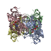
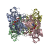
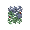
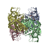
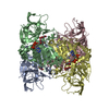
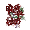
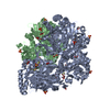
- PDB-2cda: Sulfolobus solfataricus Glucose Dehydrogenase 1 in complex with NADP -
+ Open data

- Basic information
| Entry | Database: PDB / ID: 2cda | ||||||
|---|---|---|---|---|---|---|---|
| Title | Sulfolobus solfataricus Glucose Dehydrogenase 1 in complex with NADP | ||||||
 Components Components | GLUCOSE DEHYDROGENASE | ||||||
 Keywords Keywords | OXIDOREDUCTASE / GLUCOSE DEHYDROGENASE / MEDIUM CHAIN DEHYDROGENASE FAMILY | ||||||
| Function / homology |  Function and homology information Function and homology informationaldose 1-dehydrogenase [NAD(P)+] / xylose binding / galactose catabolic process via D-galactonate / aldose 1-dehydrogenase activity / galactose 1-dehydrogenase (NADP+) / galactose 1-dehydrogenase (NADP+) activity / D-galactose 1-dehydrogenase / galactose 1-dehydrogenase activity / non-phosphorylated glucose catabolic process / glucose 1-dehydrogenase [NAD(P)+] activity ...aldose 1-dehydrogenase [NAD(P)+] / xylose binding / galactose catabolic process via D-galactonate / aldose 1-dehydrogenase activity / galactose 1-dehydrogenase (NADP+) / galactose 1-dehydrogenase (NADP+) activity / D-galactose 1-dehydrogenase / galactose 1-dehydrogenase activity / non-phosphorylated glucose catabolic process / glucose 1-dehydrogenase [NAD(P)+] activity / glucose 1-dehydrogenase (NAD+) activity / glucose 1-dehydrogenase (NADP+) activity / glucose 1-dehydrogenase [NAD(P)+] / galactose binding / D-glucose binding / NADP+ binding / NAD+ binding / protein tetramerization / zinc ion binding Similarity search - Function | ||||||
| Biological species |   SULFOLOBUS SOLFATARICUS (archaea) SULFOLOBUS SOLFATARICUS (archaea) | ||||||
| Method |  X-RAY DIFFRACTION / SYNCHROTRON / MOLECULAR REPLACEMENT / Resolution: 2.28 Å X-RAY DIFFRACTION / SYNCHROTRON / MOLECULAR REPLACEMENT / Resolution: 2.28 Å | ||||||
 Authors Authors | Milburn, C.C. / Lamble, H.J. / Theodossis, A. / Hough, D.W. / Danson, M.J. / Taylor, G.L. | ||||||
 Citation Citation | Journal: J.Biol.Chem. / Year: 2006 Title: The Structural Basis of Substrate Promiscuity in Glucose Dehydrogenase from the Hyperthermophilic Archaeon Sulfolobus Solfataricus. Authors: Milburn, C.C. / Lamble, H.J. / Theodossis, A. / Bull, S.D. / Hough, D.W. / Danson, M.J. / Taylor, G.L. | ||||||
| History |
| ||||||
| Remark 700 | SHEET THE SHEET STRUCTURE OF THIS MOLECULE IS BIFURCATED. IN ORDER TO REPRESENT THIS FEATURE IN ... SHEET THE SHEET STRUCTURE OF THIS MOLECULE IS BIFURCATED. IN ORDER TO REPRESENT THIS FEATURE IN THE SHEET RECORDS BELOW, TWO SHEETS ARE DEFINED. |
- Structure visualization
| Structure viewer | Molecule: MolmilJmol/JSmol |
|---|
- Downloads & links
-Download
| PDBx/mmCIF format | 2cda.cif.gz | 155.4 KB | Display | PDBx/mmCIF format |
|---|---|---|---|---|
| PDB format | pdb2cda.ent.gz | 123.6 KB | Display | PDB format |
| PDBx/mmJSON format | 2cda.json.gz | Tree view | PDBx/mmJSON format | |
| Others |  Other downloads Other downloads |
-Validation report
| Summary document | 2cda_validation.pdf.gz | 987.4 KB | Display | wwPDB validaton report |
|---|---|---|---|---|
| Full document | 2cda_full_validation.pdf.gz | 996.4 KB | Display | |
| Data in XML | 2cda_validation.xml.gz | 29.9 KB | Display | |
| Data in CIF | 2cda_validation.cif.gz | 41.2 KB | Display | |
| Arichive directory | https://data.pdbj.org/pub/pdb/validation_reports/cd/2cdaftp://data.pdbj.org/pub/pdb/validation_reports/cd/2cda | HTTPS FTP |
-Related structure data
| Related structure data | 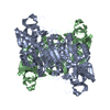 2cd9SC  2cdbC  2cdcC S: Starting model for refinement C: citing same article ( |
|---|---|
| Similar structure data |

-Links
 PDBj
PDBj


- Assembly
| Deposited unit | 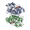
| ||||||||
|---|---|---|---|---|---|---|---|---|---|
| 1 | 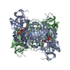
| ||||||||
| Unit cell |
|
-Components
| #1: Protein | Mass: 40945.281 Da / Num. of mol.: 2 Source method: isolated from a genetically manipulated source Source: (gene. exp.) SULFOLOBUS SOLFATARICUS (archaea) / Plasmid: PREC7 / Production host:  References: UniProt: O93715, glucose 1-dehydrogenase [NAD(P)+] #2: Chemical |   Mass: 743.405 Da / Num. of mol.: 2 / Source method: obtained synthetically / Formula: C21H28N7O17P3 Mass: 743.405 Da / Num. of mol.: 2 / Source method: obtained synthetically / Formula: C21H28N7O17P3#3: Chemical | ChemComp-ZN /   Mass: 65.409 Da / Num. of mol.: 4 / Source method: obtained synthetically / Formula: Zn Mass: 65.409 Da / Num. of mol.: 4 / Source method: obtained synthetically / Formula: Zn#4: Water | ChemComp-HOH / |  Mass: 18.015 Da / Num. of mol.: 156 / Source method: isolated from a natural source / Formula: H2O Mass: 18.015 Da / Num. of mol.: 156 / Source method: isolated from a natural source / Formula: H2OHas protein modification | Y | |
|---|
-Experimental details
-Experiment
| Experiment | Method: X-RAY DIFFRACTION / Number of used crystals: 1 |
|---|
- Sample preparation
| Crystal | Density Matthews: 2.7 Å3/Da / Density % sol: 57.4 % |
|---|---|
| Crystal grow | Details: 8 % PEG 8000, 0.1 MM TRIS (PH 8.0), 4.5 % PROPAN-2-OL |
-Data collection
| Diffraction | Mean temperature: 100 K |
|---|---|
| Diffraction source | Source: SYNCHROTRON / Site: ESRF  / Beamline: ID14-1 / Wavelength: 0.934 / Beamline: ID14-1 / Wavelength: 0.934 |
| Detector | Type: ADSC CCD / Detector: CCD / Date: Jun 12, 2004 |
| Radiation | Protocol: SINGLE WAVELENGTH / Monochromatic (M) / Laue (L): M / Scattering type: x-ray |
| Radiation wavelength | Wavelength: 0.934 Å / Relative weight: 1 |
| Reflection | Resolution: 2.28→51.2 Å / Num. obs: 38690 / % possible obs: 98.8 % / Observed criterion σ(I): 0 / Redundancy: 5.2 % / Rmerge(I) obs: 0.11 / Net I/σ(I): 12.8 |
| Reflection shell | Resolution: 2.28→2.4 Å / Redundancy: 5.2 % / Rmerge(I) obs: 0.26 / Mean I/σ(I) obs: 6.3 / % possible all: 99.6 |
- Processing
| Software |
| ||||||||||||||||||||||||||||||||||||||||||||||||||||||||||||||||||||||||||||||||||||||||||||||||||||||||||||||||||||||||||||||||||||||||||||||||||||||||||||||||||||||||||||||||||||||
|---|---|---|---|---|---|---|---|---|---|---|---|---|---|---|---|---|---|---|---|---|---|---|---|---|---|---|---|---|---|---|---|---|---|---|---|---|---|---|---|---|---|---|---|---|---|---|---|---|---|---|---|---|---|---|---|---|---|---|---|---|---|---|---|---|---|---|---|---|---|---|---|---|---|---|---|---|---|---|---|---|---|---|---|---|---|---|---|---|---|---|---|---|---|---|---|---|---|---|---|---|---|---|---|---|---|---|---|---|---|---|---|---|---|---|---|---|---|---|---|---|---|---|---|---|---|---|---|---|---|---|---|---|---|---|---|---|---|---|---|---|---|---|---|---|---|---|---|---|---|---|---|---|---|---|---|---|---|---|---|---|---|---|---|---|---|---|---|---|---|---|---|---|---|---|---|---|---|---|---|---|---|---|---|
| Refinement | Method to determine structure: MOLECULAR REPLACEMENT Starting model: PDB ENTRY 2CD9 Resolution: 2.28→138.68 Å / Cor.coef. Fo:Fc: 0.928 / Cor.coef. Fo:Fc free: 0.883 / SU B: 8.894 / SU ML: 0.16 / Cross valid method: THROUGHOUT / ESU R: 0.292 / ESU R Free: 0.227 / Stereochemistry target values: MAXIMUM LIKELIHOOD Details: HYDROGENS HAVE BEEN ADDED IN THE RIDING POSITIONS. DISORDERED LOOP REGION IS DELETED FROM THE COORDINATES
| ||||||||||||||||||||||||||||||||||||||||||||||||||||||||||||||||||||||||||||||||||||||||||||||||||||||||||||||||||||||||||||||||||||||||||||||||||||||||||||||||||||||||||||||||||||||
| Solvent computation | Ion probe radii: 0.8 Å / Shrinkage radii: 0.8 Å / VDW probe radii: 1.2 Å / Solvent model: MASK | ||||||||||||||||||||||||||||||||||||||||||||||||||||||||||||||||||||||||||||||||||||||||||||||||||||||||||||||||||||||||||||||||||||||||||||||||||||||||||||||||||||||||||||||||||||||
| Displacement parameters | Biso mean: 20.24 Å2
| ||||||||||||||||||||||||||||||||||||||||||||||||||||||||||||||||||||||||||||||||||||||||||||||||||||||||||||||||||||||||||||||||||||||||||||||||||||||||||||||||||||||||||||||||||||||
| Refinement step | Cycle: LAST / Resolution: 2.28→138.68 Å
| ||||||||||||||||||||||||||||||||||||||||||||||||||||||||||||||||||||||||||||||||||||||||||||||||||||||||||||||||||||||||||||||||||||||||||||||||||||||||||||||||||||||||||||||||||||||
| Refine LS restraints |
|