 Movie
Movie Controller

[English] 日本語
 Yorodumi
- PDB-2cah: STRUCTURE OF PROTEUS MIRABILIS PR CATALASE FOR THE NATIVE FORM (E... -
+ Open data

- Basic information
| Entry | Database: PDB / ID: 2cah | |||||||||
|---|---|---|---|---|---|---|---|---|---|---|
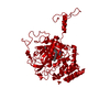
| Title | STRUCTURE OF PROTEUS MIRABILIS PR CATALASE FOR THE NATIVE FORM (E-FE(III)) COMPLEXED WITH NADPH | |||||||||
 Components Components | CATALASE | |||||||||
 Keywords Keywords | OXIDOREDUCTASE (H2O2 ACCEPTOR) / PEROXIDASE | |||||||||
| Function / homology |  Function and homology information Function and homology informationcatalase / catalase activity / hydrogen peroxide catabolic process / response to hydrogen peroxide / heme binding / metal ion binding / cytoplasm Similarity search - Function | |||||||||
| Biological species |  Proteus mirabilis (bacteria) Proteus mirabilis (bacteria) | |||||||||
| Method |  X-RAY DIFFRACTION / SYNCHROTRON / MOLECULAR REPLACEMENT / Resolution: 2.7 Å X-RAY DIFFRACTION / SYNCHROTRON / MOLECULAR REPLACEMENT / Resolution: 2.7 Å | |||||||||
 Authors Authors | Gouet, P. / Jouve, H.-M. / Dideberg, O. | |||||||||
 Citation Citation | Journal: J.Mol.Biol. / Year: 1995 Title: Crystal structure of Proteus mirabilis PR catalase with and without bound NADPH. Authors: Gouet, P. / Jouve, H.M. / Dideberg, O. #1: Journal: J.Mol.Biol. / Year: 1991Title: Crystallization and Crystal Packing of Proteus Mirabilis Pr Catalase Authors: Jouve, H.M. / Gouet, P. / Boudjada, N. / Buisson, G. / Kahn, R. / Duee, E. #2: Journal: Acta Crystallogr.,Sect.B / Year: 1986Title: The Refined Structure of Beef Liver Catalase at 2.5 Angstroms Resolution Authors: Fita, I. / Silva, A.M. / Murthy, M.R.N. / Rossmann, M.G. | |||||||||
| History |
|
- Structure visualization
| Structure viewer | Molecule: MolmilJmol/JSmol |
|---|
- Downloads & links
-Download
| PDBx/mmCIF format | 2cah.cif.gz | 116 KB | Display | PDBx/mmCIF format |
|---|---|---|---|---|
| PDB format | pdb2cah.ent.gz | 87.7 KB | Display | PDB format |
| PDBx/mmJSON format | 2cah.json.gz | Tree view | PDBx/mmJSON format | |
| Others |  Other downloads Other downloads |
-Validation report
| Arichive directory | https://data.pdbj.org/pub/pdb/validation_reports/ca/2cahftp://data.pdbj.org/pub/pdb/validation_reports/ca/2cah | HTTPS FTP |
|---|
-Related structure data


-Links
 PDBj
PDBj

- Assembly
| Deposited unit | 
| |||||||||
|---|---|---|---|---|---|---|---|---|---|---|
| 1 | 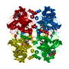
| |||||||||
| Unit cell |
| |||||||||
| Components on special symmetry positions |
| |||||||||
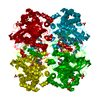
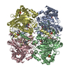
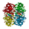
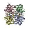
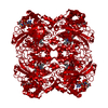
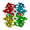
| Details | THE CATALASE OF PROTEUS MIRABILIS CRYSTALLIZES IN THE SPACE GROUP P 62 2 2 WITH ONE MONOMER PER ASYMMETRIC UNIT. THIS ENTRY GIVES THE COORDINATES OF ONE MONOMER IN THE CRYSTAL. |
-Components
| #1: Protein | Mass: 55726.348 Da / Num. of mol.: 1 / Source method: isolated from a natural source / Details: PEROXIDE RESISTANT MUTANT / Source: (natural) Proteus mirabilis (bacteria) / Organ: LIVER / References: UniProt: P42321, catalase | ||
|---|---|---|---|
| #2: Chemical | ChemComp-HEM /   Mass: 616.487 Da / Num. of mol.: 1 / Source method: obtained synthetically / Formula: C34H32FeN4O4 Mass: 616.487 Da / Num. of mol.: 1 / Source method: obtained synthetically / Formula: C34H32FeN4O4 | ||
| #3: Chemical | ChemComp-NDP /   Mass: 745.421 Da / Num. of mol.: 1 / Source method: obtained synthetically / Formula: C21H30N7O17P3 Mass: 745.421 Da / Num. of mol.: 1 / Source method: obtained synthetically / Formula: C21H30N7O17P3 | ||
| #4: Water | ChemComp-HOH /  Mass: 18.015 Da / Num. of mol.: 93 / Source method: isolated from a natural source / Formula: H2O Mass: 18.015 Da / Num. of mol.: 93 / Source method: isolated from a natural source / Formula: H2O | ||
| Compound details | THE OXYGEN OH OF THE PROXIMAL TYROSINE 337 IS DEPROTONAT| Has protein modification | Y | |
-Experimental details
-Experiment
| Experiment | Method: X-RAY DIFFRACTION / Number of used crystals: 1 |
|---|
- Sample preparation
| Crystal | Density Matthews: 4.07 Å3/Da / Density % sol: 63 % | ||||||||||||||||||||||||||||||||||||||||||||||||||||||
|---|---|---|---|---|---|---|---|---|---|---|---|---|---|---|---|---|---|---|---|---|---|---|---|---|---|---|---|---|---|---|---|---|---|---|---|---|---|---|---|---|---|---|---|---|---|---|---|---|---|---|---|---|---|---|---|
| Crystal | *PLUS | ||||||||||||||||||||||||||||||||||||||||||||||||||||||
| Crystal grow | *PLUS Temperature: 4-5 ℃ / pH: 7.5 / Method: vapor diffusion, hanging drop / Details: Jouve, H.M., (1991) J.Mol.Biol., 221, 1075. | ||||||||||||||||||||||||||||||||||||||||||||||||||||||
| Components of the solutions | *PLUS
|
-Data collection
| Diffraction source | Source: SYNCHROTRON / Site: Photon Factory  / Beamline: BL-6A / Wavelength: 1 / Beamline: BL-6A / Wavelength: 1 |
|---|---|
| Detector | Detector: FILM / Date: 1994 |
| Radiation | Monochromatic (M) / Laue (L): M / Scattering type: x-ray |
| Radiation wavelength | Wavelength: 1 Å / Relative weight: 1 |
| Reflection | Resolution: 2.7→2.823 Å / Num. obs: 24337 / % possible obs: 92 % / Observed criterion σ(I): 0 / Redundancy: 4.3 % / Rmerge(I) obs: 0.049 |
| Reflection shell | Resolution: 2.7→2.82 Å / Rsym value: 0.23 / % possible all: 92.4 |
- Processing
| Software |
| ||||||||||||||||||||||||||||||||||||||||||||||||||||||||||||
|---|---|---|---|---|---|---|---|---|---|---|---|---|---|---|---|---|---|---|---|---|---|---|---|---|---|---|---|---|---|---|---|---|---|---|---|---|---|---|---|---|---|---|---|---|---|---|---|---|---|---|---|---|---|---|---|---|---|---|---|---|---|
| Refinement | Method to determine structure: MOLECULAR REPLACEMENT Starting model: PMC WITH NADPH AT 3.1 A RESOLUTION Resolution: 2.7→15 Å / σ(F): 2 Details: NEW DATA FROM PROTEUS MIRABILIS PR CATALASE COMPLEXED WITH NADPH HAVE BEEN COLLECTED AT PHOTON FACTORY, TSUKUBA, JAPAN AND THE STRUCTURE IS NOW REFINED TO 2.7 RESOLUTION. THIS STRUCTURE IS ...Details: NEW DATA FROM PROTEUS MIRABILIS PR CATALASE COMPLEXED WITH NADPH HAVE BEEN COLLECTED AT PHOTON FACTORY, TSUKUBA, JAPAN AND THE STRUCTURE IS NOW REFINED TO 2.7 RESOLUTION. THIS STRUCTURE IS SIMILAR TO THE 3.1 A RESOLUTION STRUCTURE DESCRIBED IN REFERENCE 1 OF THIS ENTRY. REFERRING TO THIS ARTICLE, IT CAN BE NOTED THAT THE NADPH IS NOW COMPLETELY DEFINED IN THE CARDS OF ELECTRON DENSITY WHILE THE WATER MOLECULE, HOH 38, DESCRIBED AS MAYBE INVOLVED IN THE OXIDATION OF NADPH TO NADP+ IS NOT OBSERVED. WATER MOLECULES HOH 82 OF COMPOUND I STRUCTURE (2CAF) AND HOH 45 OF COMPOUND II STRUCTURE (2CAG) ARE PRESENT IN EQUIVALENT POSITION AND THE PROTEIN IS LIKELY TO BIND A WATER MOLECULE AT THIS SITE. COORDINATES FOR SIDE CHAINS OF RESIDUES 81, 204, 395, 451 AND 72, 450, 473 ARE NOT OBSERVED BEYOND CARBON CB AND CG RESPECTIVELY AND MODELED WITH AN OCCUPANCY OF 0.00 AND A TEMPERATURE FACTOR OF 99.99.
| ||||||||||||||||||||||||||||||||||||||||||||||||||||||||||||
| Displacement parameters | Biso mean: 28 Å2 | ||||||||||||||||||||||||||||||||||||||||||||||||||||||||||||
| Refinement step | Cycle: LAST / Resolution: 2.7→15 Å
| ||||||||||||||||||||||||||||||||||||||||||||||||||||||||||||
| Refine LS restraints |
| ||||||||||||||||||||||||||||||||||||||||||||||||||||||||||||
| LS refinement shell | Resolution: 2.7→2.74 Å
| ||||||||||||||||||||||||||||||||||||||||||||||||||||||||||||
| Xplor file |
|
