 Movie
Movie Controller

[English] 日本語
 Yorodumi
- PDB-2c62: Crystal Structure of the Human Transcription Cofactor PC4 in Comp... -
+ Open data

- Basic information
| Entry | Database: PDB / ID: 2c62 | ||||||
|---|---|---|---|---|---|---|---|
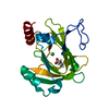
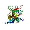
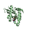
| Title | Crystal Structure of the Human Transcription Cofactor PC4 in Complex with Single-Stranded DNA | ||||||
 Components Components |
| ||||||
 Keywords Keywords | TRANSCRIPTION / TRANSCRIPTION COFACTOR / SINGLE-STRANDED DNA / PROTEIN-DNA COMPLEX / DNA UNWINDING / ACTIVATOR / DNA-BINDING / NUCLEAR PROTEIN / PHOSPHORYLATION / TRANSCRIPTION REGULATION | ||||||
| Function / homology |  Function and homology information Function and homology informationnegative regulation of DNA metabolic process / RNA polymerase II promoter clearance / positive regulation of transcription initiation by RNA polymerase II / single-stranded DNA binding / double-stranded DNA binding / transcription regulator complex / transcription coactivator activity / RNA polymerase II cis-regulatory region sequence-specific DNA binding / regulation of transcription by RNA polymerase II / nucleolus ...negative regulation of DNA metabolic process / RNA polymerase II promoter clearance / positive regulation of transcription initiation by RNA polymerase II / single-stranded DNA binding / double-stranded DNA binding / transcription regulator complex / transcription coactivator activity / RNA polymerase II cis-regulatory region sequence-specific DNA binding / regulation of transcription by RNA polymerase II / nucleolus / RNA binding / extracellular exosome / nucleoplasm / identical protein binding / nucleus Similarity search - Function | ||||||
| Biological species |  HOMO SAPIENS (human) HOMO SAPIENS (human) | ||||||
| Method |  X-RAY DIFFRACTION / SYNCHROTRON / MOLECULAR REPLACEMENT / Resolution: 1.74 Å X-RAY DIFFRACTION / SYNCHROTRON / MOLECULAR REPLACEMENT / Resolution: 1.74 Å | ||||||
 Authors Authors | Werten, S. / Moras, D. | ||||||
 Citation Citation | Journal: Nat.Struct.Mol.Biol. / Year: 2006 Title: A Global Transcription Cofactor Bound to Juxtaposed Strands of Unwound DNA Authors: Werten, S. / Moras, D. | ||||||
| History |
|
- Structure visualization
| Structure viewer | Molecule: MolmilJmol/JSmol |
|---|
- Downloads & links
-Download
| PDBx/mmCIF format | 2c62.cif.gz | 52 KB | Display | PDBx/mmCIF format |
|---|---|---|---|---|
| PDB format | pdb2c62.ent.gz | 37 KB | Display | PDB format |
| PDBx/mmJSON format | 2c62.json.gz | Tree view | PDBx/mmJSON format | |
| Others |  Other downloads Other downloads |
-Validation report
| Arichive directory | https://data.pdbj.org/pub/pdb/validation_reports/c6/2c62ftp://data.pdbj.org/pub/pdb/validation_reports/c6/2c62 | HTTPS FTP |
|---|
-Related structure data
| Related structure data |  1pcfS S: Starting model for refinement |
|---|---|
| Similar structure data |


-Links
 PDBj
PDBj


- Assembly
| Deposited unit | 
| ||||||||
|---|---|---|---|---|---|---|---|---|---|
| 1 |
| ||||||||
| Unit cell |
| ||||||||
| Details | THE BIOLOGICALLY RELEVANT STATE OF THE MOLECULE ISTHE CONTENTS OF THE ASYMMETRIC UNIT. |
-Components
| #1: Protein | Mass: 7784.024 Da / Num. of mol.: 2 / Fragment: C-TERMINAL SSDNA-BINDING DOMAIN, RESIDUES 62-126 Source method: isolated from a genetically manipulated source Details: N-TERMINAL ALA RESIDUE RESULTS FROM VECTOR SEQUENCE Source: (gene. exp.) HOMO SAPIENS (human) / Plasmid: PET-11A / Production host:  #2: DNA chain | | Mass: 6063.912 Da / Num. of mol.: 1 / Source method: obtained synthetically #3: Chemical |   Mass: 96.063 Da / Num. of mol.: 2 / Source method: obtained synthetically / Formula: SO4 Mass: 96.063 Da / Num. of mol.: 2 / Source method: obtained synthetically / Formula: SO4#4: Water | ChemComp-HOH / |  Mass: 18.015 Da / Num. of mol.: 176 / Source method: isolated from a natural source / Formula: H2O Mass: 18.015 Da / Num. of mol.: 176 / Source method: isolated from a natural source / Formula: H2OCompound details | GENERAL COACTIVATOR THAT FUNCTIONS IN COOPERATION WITH TAFS AND MEDIATES FUNCTIONAL INTERACTIONS ...GENERAL COACTIVATO | Sequence details | C-TERMINAL DOMAIN ONLY, PRECEEDED BY ALA FROM EXPRESSION | |
|---|
-Experimental details
-Experiment
| Experiment | Method: X-RAY DIFFRACTION / Number of used crystals: 1 |
|---|
- Sample preparation
| Crystal | Density Matthews: 3.33 Å3/Da / Density % sol: 62.77 % |
|---|---|
| Crystal grow | pH: 6.75 / Details: 0.1 M ADA PH 6.75, 1.9 M AMMONIUM SULFATE |
-Data collection
| Diffraction | Mean temperature: 100 K |
|---|---|
| Diffraction source | Source: SYNCHROTRON / Site: ESRF  / Beamline: ID14-4 / Wavelength: 0.9793 / Beamline: ID14-4 / Wavelength: 0.9793 |
| Detector | Type: ADSC CCD / Detector: CCD / Date: Jun 20, 2003 / Details: TOROIDAL FOCUSING MIRROR |
| Radiation | Monochromator: DOUBLE CRYSTAL MONOCHROMATOR / Protocol: SINGLE WAVELENGTH / Monochromatic (M) / Laue (L): M / Scattering type: x-ray |
| Radiation wavelength | Wavelength: 0.9793 Å / Relative weight: 1 |
| Reflection | Resolution: 1.74→20 Å / Num. obs: 31658 / % possible obs: 99.9 % / Observed criterion σ(I): 0 / Redundancy: 6.3 % / Biso Wilson estimate: 29.9 Å2 / Rmerge(I) obs: 0.04 / Net I/σ(I): 21.3 |
| Reflection shell | Resolution: 1.74→1.8 Å / Redundancy: 4.8 % / Rmerge(I) obs: 0.3 / Mean I/σ(I) obs: 5.43 / % possible all: 100 |
- Processing
| Software |
| ||||||||||||||||||||||||||||||||||||||||||||||||||||||||||||||||||||||||||||||||
|---|---|---|---|---|---|---|---|---|---|---|---|---|---|---|---|---|---|---|---|---|---|---|---|---|---|---|---|---|---|---|---|---|---|---|---|---|---|---|---|---|---|---|---|---|---|---|---|---|---|---|---|---|---|---|---|---|---|---|---|---|---|---|---|---|---|---|---|---|---|---|---|---|---|---|---|---|---|---|---|---|---|
| Refinement | Method to determine structure: MOLECULAR REPLACEMENT Starting model: PDB ENTRY 1PCF, CHAINS A AND B Resolution: 1.74→19.97 Å / Rfactor Rfree error: 0.006 / Data cutoff high absF: 1346476 / Isotropic thermal model: RESTRAINED / Cross valid method: THROUGHOUT / σ(F): 0 / Stereochemistry target values: MAXIMUM LIKELIHOOD
| ||||||||||||||||||||||||||||||||||||||||||||||||||||||||||||||||||||||||||||||||
| Solvent computation | Solvent model: FLAT MODEL / Bsol: 52.429 Å2 / ksol: 0.363232 e/Å3 | ||||||||||||||||||||||||||||||||||||||||||||||||||||||||||||||||||||||||||||||||
| Displacement parameters | Biso mean: 42.1 Å2
| ||||||||||||||||||||||||||||||||||||||||||||||||||||||||||||||||||||||||||||||||
| Refine analyze |
| ||||||||||||||||||||||||||||||||||||||||||||||||||||||||||||||||||||||||||||||||
| Refinement step | Cycle: LAST / Resolution: 1.74→19.97 Å
| ||||||||||||||||||||||||||||||||||||||||||||||||||||||||||||||||||||||||||||||||
| Refine LS restraints |
| ||||||||||||||||||||||||||||||||||||||||||||||||||||||||||||||||||||||||||||||||
| LS refinement shell | Resolution: 1.74→1.8 Å / Rfactor Rfree error: 0.032 / Total num. of bins used: 10
| ||||||||||||||||||||||||||||||||||||||||||||||||||||||||||||||||||||||||||||||||
| Xplor file |
|
