 Movie
Movie Controller

[English] 日本語
 Yorodumi
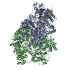
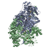
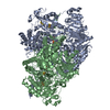
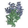
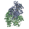
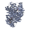
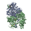
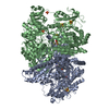
- PDB-2c3u: Crystal Structure Of Pyruvate-Ferredoxin Oxidoreductase From Desu... -
+ Open data

- Basic information
| Entry | Database: PDB / ID: 2c3u | |||||||||
|---|---|---|---|---|---|---|---|---|---|---|
| Title | Crystal Structure Of Pyruvate-Ferredoxin Oxidoreductase From Desulfovibrio africanus, Oxygen inhibited form | |||||||||
 Components Components | PYRUVATE-FERREDOXIN OXIDOREDUCTASE | |||||||||
 Keywords Keywords | OXIDOREDUCTASE / 4FE-4S / IRON / IRON-SULFUR / IRON-SULFUR CLUSTER / PYRUVATE CATABOLISM / TPP-DEPENDENT ENZYME / METAL-BINDING / ELECTRON TRANSPORT | |||||||||
| Function / homology |  Function and homology information Function and homology informationpyruvate synthase (ferredoxin) / pyruvate synthase activity / thiamine pyrophosphate binding / electron transport chain / 4 iron, 4 sulfur cluster binding / response to oxidative stress / iron ion binding / cytoplasm Similarity search - Function | |||||||||
| Biological species |  DESULFOVIBRIO AFRICANUS (bacteria) DESULFOVIBRIO AFRICANUS (bacteria) | |||||||||
| Method |  X-RAY DIFFRACTION / SYNCHROTRON / MOLECULAR REPLACEMENT / Resolution: 2.32 Å X-RAY DIFFRACTION / SYNCHROTRON / MOLECULAR REPLACEMENT / Resolution: 2.32 Å | |||||||||
 Authors Authors | Cavazza, C. / Contreras-Martel, C. / Pieulle, L. / Chabriere, E. / Hatchikian, E.C. / Fontecilla-Camps, J.C. | |||||||||
 Citation Citation | Journal: Structure / Year: 2006 Title: Flexibility of Thiamine Diphosphate Revealed by Kinetic Crystallographic Studies of the Reaction of Pyruvate-Ferredoxin Oxidoreductase with Pyruvate. Authors: Cavazza, C. / Contreras-Martel, C. / Pieulle, L. / Chabriere, E. / Hatchikian, E.C. / Fontecilla-Camps, J.C. #1: Journal: Science / Year: 2001Title: Crystal Structure of the Free Radical Intermediate of Pyruvate:Ferredoxin Oxidoreductase. Authors: Chabriere, E. / Vernede, X. / Guigliarelli, B. / Charon, M.-H. / Hatchikian, E.C. / Fontecilla-Camps, J.C. | |||||||||
| History |
|
- Structure visualization
| Structure viewer | Molecule: MolmilJmol/JSmol |
|---|
- Downloads & links
-Download
| PDBx/mmCIF format | 2c3u.cif.gz | 497.5 KB | Display | PDBx/mmCIF format |
|---|---|---|---|---|
| PDB format | pdb2c3u.ent.gz | 396.9 KB | Display | PDB format |
| PDBx/mmJSON format | 2c3u.json.gz | Tree view | PDBx/mmJSON format | |
| Others |  Other downloads Other downloads |
-Validation report
| Arichive directory | https://data.pdbj.org/pub/pdb/validation_reports/c3/2c3uftp://data.pdbj.org/pub/pdb/validation_reports/c3/2c3u | HTTPS FTP |
|---|
-Related structure data
| Related structure data |  2c3mC  2c3oC 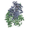 2c3pC  2c3yC  2c42C  2uzaC  1kekS S: Starting model for refinement C: citing same article ( |
|---|---|
| Similar structure data |


-Links
 PDBj
PDBj


- Assembly
| Deposited unit | 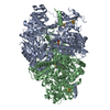
| ||||||||
|---|---|---|---|---|---|---|---|---|---|
| 1 |
| ||||||||
| Unit cell |
|
-Components
-Protein , 1 types, 2 molecules AB
| #1: Protein | Mass: 133703.906 Da / Num. of mol.: 2 / Source method: isolated from a natural source Details: COMPLEXED WITH IRON/SULFUR CLUSTER, THIAMIN DIPHOSPHATE. CRYSTAL SOAKED OVERNIGHT WITH PYRUVATE Source: (natural) DESULFOVIBRIO AFRICANUS (bacteria) / References: UniProt: P94692, pyruvate synthase (ferredoxin) |
|---|
-Non-polymers , 6 types, 776 molecules 
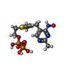


| #2: Chemical | ChemComp-SF4 /  Mass: 351.640 Da / Num. of mol.: 6 / Source method: obtained synthetically / Formula: Fe4S4 Mass: 351.640 Da / Num. of mol.: 6 / Source method: obtained synthetically / Formula: Fe4S4#3: Chemical |  Mass: 441.314 Da / Num. of mol.: 2 / Source method: obtained synthetically / Formula: C12H19N4O8P2S Mass: 441.314 Da / Num. of mol.: 2 / Source method: obtained synthetically / Formula: C12H19N4O8P2S#4: Chemical |  Mass: 88.062 Da / Num. of mol.: 2 / Source method: obtained synthetically / Formula: C3H4O3 Mass: 88.062 Da / Num. of mol.: 2 / Source method: obtained synthetically / Formula: C3H4O3#5: Chemical |  Mass: 24.305 Da / Num. of mol.: 2 / Source method: obtained synthetically / Formula: Mg Mass: 24.305 Da / Num. of mol.: 2 / Source method: obtained synthetically / Formula: Mg#6: Chemical |  Mass: 40.078 Da / Num. of mol.: 2 / Source method: obtained synthetically / Formula: Ca Mass: 40.078 Da / Num. of mol.: 2 / Source method: obtained synthetically / Formula: Ca#7: Water | ChemComp-HOH / | Mass: 18.015 Da / Num. of mol.: 762 / Source method: isolated from a natural source / Formula: H2O |
|---|
-Details
| Has protein modification | Y |
|---|
-Experimental details
-Experiment
| Experiment | Method: X-RAY DIFFRACTION / Number of used crystals: 1 |
|---|
- Sample preparation
| Crystal | Density Matthews: 2.52 Å3/Da / Density % sol: 49.32 % |
|---|---|
| Crystal grow | pH: 9 / Details: 10% PEG6000, 100MM MGCL2, 100MM TRIS-HCL PH 9 |
-Data collection
| Diffraction | Mean temperature: 100 K |
|---|---|
| Diffraction source | Source: SYNCHROTRON / Site: ESRF  / Beamline: ID14-2 / Wavelength: 0.933 / Beamline: ID14-2 / Wavelength: 0.933 |
| Detector | Type: ADSC CCD / Detector: CCD / Date: Apr 15, 2001 |
| Radiation | Protocol: SINGLE WAVELENGTH / Monochromatic (M) / Laue (L): M / Scattering type: x-ray |
| Radiation wavelength | Wavelength: 0.933 Å / Relative weight: 1 |
| Reflection | Resolution: 2.32→55 Å / Num. obs: 112651 / % possible obs: 95.9 % / Observed criterion σ(I): 2 / Redundancy: 3.5 % / Biso Wilson estimate: 13.3 Å2 / Rsym value: 0.1 / Net I/σ(I): 11.63 |
| Reflection shell | Resolution: 2.32→2.5 Å / Redundancy: 3.5 % / Mean I/σ(I) obs: 4.96 / Rsym value: 0.28 / % possible all: 90.3 |
- Processing
| Software |
| ||||||||||||||||||||||||||||||||||||||||||||||||||||||||||||||||||||||||||||||||
|---|---|---|---|---|---|---|---|---|---|---|---|---|---|---|---|---|---|---|---|---|---|---|---|---|---|---|---|---|---|---|---|---|---|---|---|---|---|---|---|---|---|---|---|---|---|---|---|---|---|---|---|---|---|---|---|---|---|---|---|---|---|---|---|---|---|---|---|---|---|---|---|---|---|---|---|---|---|---|---|---|---|
| Refinement | Method to determine structure: MOLECULAR REPLACEMENT Starting model: PDB ENTRY 1KEK Resolution: 2.32→49.48 Å / Rfactor Rfree error: 0.003 / Data cutoff high absF: 3866757.12 / Isotropic thermal model: RESTRAINED / Cross valid method: THROUGHOUT / σ(F): 0
| ||||||||||||||||||||||||||||||||||||||||||||||||||||||||||||||||||||||||||||||||
| Solvent computation | Solvent model: FLAT MODEL / Bsol: 27.3221 Å2 / ksol: 0.344666 e/Å3 | ||||||||||||||||||||||||||||||||||||||||||||||||||||||||||||||||||||||||||||||||
| Displacement parameters | Biso mean: 23.2 Å2
| ||||||||||||||||||||||||||||||||||||||||||||||||||||||||||||||||||||||||||||||||
| Refine analyze |
| ||||||||||||||||||||||||||||||||||||||||||||||||||||||||||||||||||||||||||||||||
| Refinement step | Cycle: LAST / Resolution: 2.32→49.48 Å
| ||||||||||||||||||||||||||||||||||||||||||||||||||||||||||||||||||||||||||||||||
| Refine LS restraints |
| ||||||||||||||||||||||||||||||||||||||||||||||||||||||||||||||||||||||||||||||||
| LS refinement shell | Resolution: 2.32→2.47 Å / Rfactor Rfree error: 0.01 / Total num. of bins used: 6
| ||||||||||||||||||||||||||||||||||||||||||||||||||||||||||||||||||||||||||||||||
| Xplor file |
|
