Resolution: 1.87→1.97 Å / Mean I/σ(I) obs: 1.4 / Rsym value: 0.293 / % possible all: 97.3
Reflection
*PLUS
Highest resolution: 1.87 Å / Rmerge(I) obs: 0.084
Reflection shell
*PLUS
% possible obs: 97.3 % / Rmerge(I) obs: 0.293
-
Processing
Software
Name
Version
Classification
MOSFLM
datareduction
SCALA
datascaling
X-PLOR
modelbuilding
X-PLOR
refinement
REFMAC
refinement
CCP4
(SCALA)
datascaling
X-PLOR
phasing
Refinement
Method to determine structure: MOLECULAR REPLACEMENT / Resolution: 1.9→27.38 Å / Rfactor Rfree error: 0.002 / Data cutoff high absF: 3963562.61 / Data cutoff low absF: 0 / Isotropic thermal model: RESTRAINED / Cross valid method: THROUGHOUT / σ(F): 0 / Stereochemistry target values: X-PLOR + Home dictionary Details: THIS REFINED SET DIFFERS SLIGHTLY FROM THE ONE REPORTED IN REF. FOR EXAMPLE, THE C2ALPHA-C2 DISTANCES OF THE RADICAL SPECIES DO NOT MATCH EXACTLY THE PUBLISHED ONES.
In the structure databanks used in Yorodumi, some data are registered as the other names, "COVID-19 virus" and "2019-nCoV". Here are the details of the virus and the list of structure data.
Jan 31, 2019. EMDB accession codes are about to change! (news from PDBe EMDB page)
EMDB accession codes are about to change! (news from PDBe EMDB page)
The allocation of 4 digits for EMDB accession codes will soon come to an end. Whilst these codes will remain in use, new EMDB accession codes will include an additional digit and will expand incrementally as the available range of codes is exhausted. The current 4-digit format prefixed with “EMD-” (i.e. EMD-XXXX) will advance to a 5-digit format (i.e. EMD-XXXXX), and so on. It is currently estimated that the 4-digit codes will be depleted around Spring 2019, at which point the 5-digit format will come into force.
The EM Navigator/Yorodumi systems omit the EMD- prefix.
Related info.:Q: What is EMD? / ID/Accession-code notation in Yorodumi/EM Navigator
Yorodumi is a browser for structure data from EMDB, PDB, SASBDB, etc.
This page is also the successor to EM Navigator detail page, and also detail information page/front-end page for Omokage search.
The word "yorodu" (or yorozu) is an old Japanese word meaning "ten thousand". "mi" (miru) is to see.
Related info.:EMDB / PDB / SASBDB / Comparison of 3 databanks / Yorodumi Search / Aug 31, 2016. New EM Navigator & Yorodumi / Yorodumi Papers / Jmol/JSmol / Function and homology information / Changes in new EM Navigator and Yorodumi
 Movie
Movie Controller

 Yorodumi
 Open data
 Basic information
 Components
 Keywords
 Function and homology information
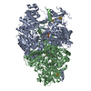
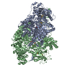
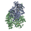
 Desulfovibrio africanus (bacteria)
 X-RAY DIFFRACTION /
  Authors
 Citation
 Structure visualization
 Downloads & links
 Other downloads
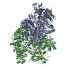


 PDBj
PDBj


 Assembly


 Mass: 24.305 Da / Num. of mol.: 2 / Source method: obtained synthetically / Formula: Mg
 Mass: 40.078 Da / Num. of mol.: 2 / Source method: obtained synthetically / Formula: Ca
 Mass: 351.640 Da / Num. of mol.: 6 / Source method: obtained synthetically / Formula: Fe4S4
 Mass: 467.351 Da / Num. of mol.: 2 / Source method: obtained synthetically / Formula: C14H21N4O8P2S
 Mass: 44.010 Da / Num. of mol.: 2 / Source method: obtained synthetically / Formula: CO2
 Sample preparation
 / Beamline: ID14-3 / Wavelength: 1.005 Å
 Processing