 Movie
Movie Controller

[English] 日本語
 Yorodumi
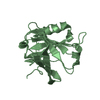
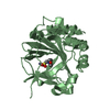
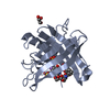
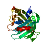
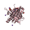
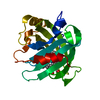
- PDB-2c2p: The crystal structure of mismatch specific uracil-DNA glycosylase... -
+ Open data

- Basic information
| Entry | Database: PDB / ID: 2c2p | ||||||
|---|---|---|---|---|---|---|---|
| Title | The crystal structure of mismatch specific uracil-DNA glycosylase (MUG) from Deinococcus radiodurans | ||||||
 Components Components | G/U MISMATCH-SPECIFIC DNA GLYCOSYLASE | ||||||
 Keywords Keywords | HYDROLASE / DEINOCOCCUS RADIODURANS / RADIATION RESISTANCE / DNA REPAIR ENZYMES / URACIL-DNA GLYCOSYLASE / MISMATCH SPECIFIC DNA-GLYCOSYLASE / MUG | ||||||
| Function / homology |  Function and homology information Function and homology informationpyrimidine-specific mismatch base pair DNA N-glycosylase activity / base-excision repair, AP site formation / uracil DNA N-glycosylase activity Similarity search - Function | ||||||
| Biological species |  DEINOCOCCUS RADIODURANS (radioresistant) DEINOCOCCUS RADIODURANS (radioresistant) | ||||||
| Method |  X-RAY DIFFRACTION / SYNCHROTRON / MOLECULAR REPLACEMENT / Resolution: 1.75 Å X-RAY DIFFRACTION / SYNCHROTRON / MOLECULAR REPLACEMENT / Resolution: 1.75 Å | ||||||
 Authors Authors | Moe, E. / Leiros, I. / Smalas, A.O. / McSweeney, S. | ||||||
 Citation Citation | Journal: J.Biol.Chem. / Year: 2006 Title: The Crystal Structure of Mismatch Specific Uracil-DNA Glycosylase (Mug) from Deinococcus Radiodurans Reveals a Novel Catalytic Residue and Broad Substrate Specificity Authors: Moe, E. / Leiros, I. / Smalas, A.O. / Mcsweeney, S. | ||||||
| History |
|
- Structure visualization
| Structure viewer | Molecule: MolmilJmol/JSmol |
|---|
- Downloads & links
-Download
| PDBx/mmCIF format | 2c2p.cif.gz | 52.8 KB | Display | PDBx/mmCIF format |
|---|---|---|---|---|
| PDB format | pdb2c2p.ent.gz | 37.4 KB | Display | PDB format |
| PDBx/mmJSON format | 2c2p.json.gz | Tree view | PDBx/mmJSON format | |
| Others |  Other downloads Other downloads |
-Validation report
| Summary document | 2c2p_validation.pdf.gz | 440.1 KB | Display | wwPDB validaton report |
|---|---|---|---|---|
| Full document | 2c2p_full_validation.pdf.gz | 442.3 KB | Display | |
| Data in XML | 2c2p_validation.xml.gz | 11.1 KB | Display | |
| Data in CIF | 2c2p_validation.cif.gz | 15.4 KB | Display | |
| Arichive directory | https://data.pdbj.org/pub/pdb/validation_reports/c2/2c2pftp://data.pdbj.org/pub/pdb/validation_reports/c2/2c2p | HTTPS FTP |
-Related structure data
| Related structure data | 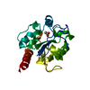 2c2qC 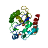 1mugS C: citing same article ( S: Starting model for refinement |
|---|---|
| Similar structure data |


-Links
 PDBj
PDBj- Assembly
| Deposited unit | 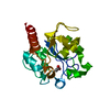
| ||||||||
|---|---|---|---|---|---|---|---|---|---|
| 1 |
| ||||||||
| Unit cell |
| ||||||||
| Components on special symmetry positions |
|
-Components
| #1: Protein | Mass: 21785.584 Da / Num. of mol.: 1 Source method: isolated from a genetically manipulated source Details: ACETATE BOUND IN THE SUBSTRATE POCKET Source: (gene. exp.) DEINOCOCCUS RADIODURANS (radioresistant)Strain: R1 / Production host: References: UniProt: Q9RWF4, Hydrolases; Glycosylases; Hydrolysing N-glycosyl compounds |
|---|---|
| #2: Chemical | ChemComp-ACT /   Mass: 59.044 Da / Num. of mol.: 1 / Source method: obtained synthetically / Formula: C2H3O2 Mass: 59.044 Da / Num. of mol.: 1 / Source method: obtained synthetically / Formula: C2H3O2 |
| #3: Water | ChemComp-HOH /  Mass: 18.015 Da / Num. of mol.: 152 / Source method: isolated from a natural source / Formula: H2O Mass: 18.015 Da / Num. of mol.: 152 / Source method: isolated from a natural source / Formula: H2O |
-Experimental details
-Experiment
| Experiment | Method: X-RAY DIFFRACTION / Number of used crystals: 1 |
|---|
- Sample preparation
| Crystal | Density Matthews: 2.6 Å3/Da / Density % sol: 51.8 % |
|---|---|
| Crystal grow | pH: 6.5 Details: 0.2 M SODIUM ACETATE TRIHYDRATE, 0.1 M SODIUM CACODYLATE PH 6.5, 30% W/V PEG 8000 |
-Data collection
| Diffraction | Mean temperature: 100 K |
|---|---|
| Diffraction source | Source: SYNCHROTRON / Site: ESRF  / Beamline: ID14-4 / Wavelength: 0.939272 / Beamline: ID14-4 / Wavelength: 0.939272 |
| Detector | Type: ADSC CCD / Detector: CCD / Date: Oct 6, 2003 |
| Radiation | Protocol: SINGLE WAVELENGTH / Monochromatic (M) / Laue (L): M / Scattering type: x-ray |
| Radiation wavelength | Wavelength: 0.939272 Å / Relative weight: 1 |
| Reflection | Resolution: 1.75→12 Å / Num. obs: 22686 / % possible obs: 99.9 % / Observed criterion σ(I): 0 / Redundancy: 5.3 % / Rmerge(I) obs: 0.05 / Net I/σ(I): 18.8 |
| Reflection shell | Resolution: 1.75→1.84 Å / Redundancy: 5.1 % / Rmerge(I) obs: 0.53 / Mean I/σ(I) obs: 2.8 / % possible all: 100 |
- Processing
| Software |
| ||||||||||||||||||||||||||||||||||||||||||||||||||||||||||||||||||||||||||||||||||||||||||||||||||||||||||||||||||||||||||||||||||||||||||||||||||||||||||||||||||||||||||||||||||||||
|---|---|---|---|---|---|---|---|---|---|---|---|---|---|---|---|---|---|---|---|---|---|---|---|---|---|---|---|---|---|---|---|---|---|---|---|---|---|---|---|---|---|---|---|---|---|---|---|---|---|---|---|---|---|---|---|---|---|---|---|---|---|---|---|---|---|---|---|---|---|---|---|---|---|---|---|---|---|---|---|---|---|---|---|---|---|---|---|---|---|---|---|---|---|---|---|---|---|---|---|---|---|---|---|---|---|---|---|---|---|---|---|---|---|---|---|---|---|---|---|---|---|---|---|---|---|---|---|---|---|---|---|---|---|---|---|---|---|---|---|---|---|---|---|---|---|---|---|---|---|---|---|---|---|---|---|---|---|---|---|---|---|---|---|---|---|---|---|---|---|---|---|---|---|---|---|---|---|---|---|---|---|---|---|
| Refinement | Method to determine structure: MOLECULAR REPLACEMENT Starting model: PDB ENTRY 1MUG Resolution: 1.75→87.71 Å / Cor.coef. Fo:Fc: 0.962 / Cor.coef. Fo:Fc free: 0.941 / SU B: 2.32 / SU ML: 0.074 / Cross valid method: THROUGHOUT / ESU R: 0.108 / ESU R Free: 0.11 / Stereochemistry target values: MAXIMUM LIKELIHOOD Details: HYDROGENS HAVE BEEN ADDED IN THE RIDING POSITIONS. RESIDUES 1-6 AND 190-199 WERE DISORDERED AND THEREFORE NOT MODELLED.
| ||||||||||||||||||||||||||||||||||||||||||||||||||||||||||||||||||||||||||||||||||||||||||||||||||||||||||||||||||||||||||||||||||||||||||||||||||||||||||||||||||||||||||||||||||||||
| Solvent computation | Ion probe radii: 0.8 Å / Shrinkage radii: 0.8 Å / VDW probe radii: 1.4 Å / Solvent model: BABINET MODEL WITH MASK | ||||||||||||||||||||||||||||||||||||||||||||||||||||||||||||||||||||||||||||||||||||||||||||||||||||||||||||||||||||||||||||||||||||||||||||||||||||||||||||||||||||||||||||||||||||||
| Displacement parameters | Biso mean: 23.13 Å2
| ||||||||||||||||||||||||||||||||||||||||||||||||||||||||||||||||||||||||||||||||||||||||||||||||||||||||||||||||||||||||||||||||||||||||||||||||||||||||||||||||||||||||||||||||||||||
| Refinement step | Cycle: LAST / Resolution: 1.75→87.71 Å
| ||||||||||||||||||||||||||||||||||||||||||||||||||||||||||||||||||||||||||||||||||||||||||||||||||||||||||||||||||||||||||||||||||||||||||||||||||||||||||||||||||||||||||||||||||||||
| Refine LS restraints |
|