 Movie
Movie Controller

[English] 日本語
 Yorodumi
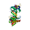
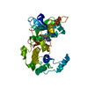
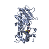
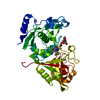
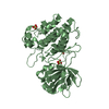
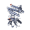
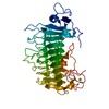
- PDB-2c27: The Structure of Mycothiol Synthase in Complex with des- AcetylMy... -
+ Open data

- Basic information
| Entry | Database: PDB / ID: 2c27 | |||||||||
|---|---|---|---|---|---|---|---|---|---|---|
| Title | The Structure of Mycothiol Synthase in Complex with des- AcetylMycothiol and CoenzymeA. | |||||||||
 Components Components | MYCOTHIOL SYNTHASE | |||||||||
 Keywords Keywords | SYNTHASE / MYCOBACTERIUM TUBERCULOSIS / MYCOTHIOL SYNTHASE / DES- ACETYLMYCOTHIOL / ACETYLTRANSFERASE / GNAT / GCN5 RELATED N- ACETYLTRANSFERASE | |||||||||
| Function / homology |  Function and homology information Function and homology informationmycothiol synthase / mycothiol synthase activity / : / mycothiol metabolic process / Mycothiol biosynthesis / mycothiol biosynthetic process / protein-N-terminal-alanine acetyltransferase activity / biological process involved in interaction with host / acyltransferase activity, transferring groups other than amino-acyl groups / cellular response to acidic pH ...mycothiol synthase / mycothiol synthase activity / : / mycothiol metabolic process / Mycothiol biosynthesis / mycothiol biosynthetic process / protein-N-terminal-alanine acetyltransferase activity / biological process involved in interaction with host / acyltransferase activity, transferring groups other than amino-acyl groups / cellular response to acidic pH / cellular response to hydrogen peroxide / cytosol Similarity search - Function | |||||||||
| Biological species |   MYCOBACTERIUM TUBERCULOSIS (bacteria) MYCOBACTERIUM TUBERCULOSIS (bacteria) | |||||||||
| Method |  X-RAY DIFFRACTION / MOLECULAR REPLACEMENT / Resolution: 1.8 Å X-RAY DIFFRACTION / MOLECULAR REPLACEMENT / Resolution: 1.8 Å | |||||||||
 Authors Authors | Vetting, M.W. / Yu, M. / Rendle, P.M. / Blanchard, J.S. | |||||||||
 Citation Citation | Journal: J.Biol.Chem. / Year: 2006 Title: The Substrate-Induced Conformational Change of Mycobacterium Tuberculosis Mycothiol Synthase. Authors: Vetting, M.W. / Yu, M. / Rendle, P.M. / Blanchard, J.S. | |||||||||
| History |
|
- Structure visualization
| Structure viewer | Molecule: MolmilJmol/JSmol |
|---|
- Downloads & links
-Download
| PDBx/mmCIF format | 2c27.cif.gz | 78.9 KB | Display | PDBx/mmCIF format |
|---|---|---|---|---|
| PDB format | pdb2c27.ent.gz | 56.2 KB | Display | PDB format |
| PDBx/mmJSON format | 2c27.json.gz | Tree view | PDBx/mmJSON format | |
| Others |  Other downloads Other downloads |
-Validation report
| Arichive directory | https://data.pdbj.org/pub/pdb/validation_reports/c2/2c27ftp://data.pdbj.org/pub/pdb/validation_reports/c2/2c27 | HTTPS FTP |
|---|
-Related structure data
| Related structure data | 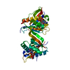 1ozpS S: Starting model for refinement |
|---|---|
| Similar structure data |


-Links
 PDBj
PDBj


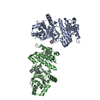
- Assembly
| Deposited unit | 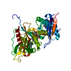
| ||||||||
|---|---|---|---|---|---|---|---|---|---|
| 1 |
| ||||||||
| Unit cell |
|
-Components
| #1: Protein | Mass: 33639.043 Da / Num. of mol.: 1 Source method: isolated from a genetically manipulated source Source: (gene. exp.) MYCOBACTERIUM TUBERCULOSIS (bacteria) / Strain: H37RV / Plasmid: PET28A / Production host: |
|---|---|
| #2: Sugar | ChemComp-MA8 / (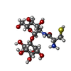  Type: D-saccharide / Mass: 444.455 Da / Num. of mol.: 1 / Source method: obtained synthetically / Formula: C15H28N2O11S Type: D-saccharide / Mass: 444.455 Da / Num. of mol.: 1 / Source method: obtained synthetically / Formula: C15H28N2O11S |
| #3: Chemical | ChemComp-ACO /   Mass: 809.571 Da / Num. of mol.: 1 / Source method: obtained synthetically / Formula: C23H38N7O17P3S Mass: 809.571 Da / Num. of mol.: 1 / Source method: obtained synthetically / Formula: C23H38N7O17P3S |
| #4: Chemical | ChemComp-COA /   Mass: 767.534 Da / Num. of mol.: 1 / Source method: obtained synthetically / Formula: C21H36N7O16P3S Mass: 767.534 Da / Num. of mol.: 1 / Source method: obtained synthetically / Formula: C21H36N7O16P3S |
| #5: Water | ChemComp-HOH /  Mass: 18.015 Da / Num. of mol.: 175 / Source method: isolated from a natural source / Formula: H2O Mass: 18.015 Da / Num. of mol.: 175 / Source method: isolated from a natural source / Formula: H2O |
| Compound details | DES-ACETYLMYCO |
-Experimental details
-Experiment
| Experiment | Method: X-RAY DIFFRACTION / Number of used crystals: 1 |
|---|
- Sample preparation
| Crystal | Density Matthews: 1.93 Å3/Da / Density % sol: 35.67 % |
|---|---|
| Crystal grow | pH: 8.5 Details: 1 M NA3CITRATE PH 8.5 100 MM BICINE PH 8.8 7.7 MM DESACETYL-MYCOTHIOL 5.3 MM COENZYMEA 40 MM DTT |
-Data collection
| Diffraction | Mean temperature: 292 K |
|---|---|
| Diffraction source | Source: ROTATING ANODE / Type: RIGAKU RU200B / Wavelength: 1.5418 |
| Detector | Type: RIGAKU RAXIS-IV / Detector: IMAGE PLATE / Details: OSMIC CONFOCAL MAXFLUX OPTICS |
| Radiation | Protocol: SINGLE WAVELENGTH / Monochromatic (M) / Laue (L): M / Scattering type: x-ray |
| Radiation wavelength | Wavelength: 1.5418 Å / Relative weight: 1 |
| Reflection | Resolution: 1.8→30 Å / Num. obs: 24682 / % possible obs: 96.7 % / Observed criterion σ(I): 0 / Redundancy: 2.9 % / Rmerge(I) obs: 0.09 / Net I/σ(I): 14.2 |
| Reflection shell | Resolution: 1.8→1.86 Å / Redundancy: 2.7 % / Rmerge(I) obs: 0.14 / Mean I/σ(I) obs: 7.6 / % possible all: 94.3 |
- Processing
| Software |
| ||||||||||||||||||||||||||||||||||||||||||||||||||||||||||||
|---|---|---|---|---|---|---|---|---|---|---|---|---|---|---|---|---|---|---|---|---|---|---|---|---|---|---|---|---|---|---|---|---|---|---|---|---|---|---|---|---|---|---|---|---|---|---|---|---|---|---|---|---|---|---|---|---|---|---|---|---|---|
| Refinement | Method to determine structure: MOLECULAR REPLACEMENT Starting model: PDB ENTRY 1OZP Resolution: 1.8→30 Å / Cross valid method: THROUGHOUT / σ(F): 0 / Stereochemistry target values: MAXIMUM LIKELIHOOD
| ||||||||||||||||||||||||||||||||||||||||||||||||||||||||||||
| Solvent computation | Solvent model: FLAT MODEL / Bsol: 58.7512 Å2 / ksol: 0.407886 e/Å3 | ||||||||||||||||||||||||||||||||||||||||||||||||||||||||||||
| Displacement parameters | Biso mean: 23.6 Å2
| ||||||||||||||||||||||||||||||||||||||||||||||||||||||||||||
| Refinement step | Cycle: LAST / Resolution: 1.8→30 Å
| ||||||||||||||||||||||||||||||||||||||||||||||||||||||||||||
| Refine LS restraints |
| ||||||||||||||||||||||||||||||||||||||||||||||||||||||||||||
| LS refinement shell | Resolution: 1.8→1.88 Å / Total num. of bins used: 10 /
|
