 Movie
Movie Controller

[English] 日本語
 Yorodumi
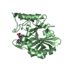
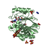
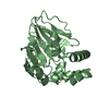
- PDB-2c1f: The structure of the family 11 xylanase from Neocallimastix patri... -
+ Open data

- Basic information
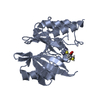
| Entry | Database: PDB / ID: 2c1f | ||||||
|---|---|---|---|---|---|---|---|
| Title | The structure of the family 11 xylanase from Neocallimastix patriciarum | ||||||
 Components Components | BIFUNCTIONAL ENDO-1,4-BETA-XYLANASE A | ||||||
 Keywords Keywords | HYDROLASE / GLYCOSIDASE | ||||||
| Function / homology |  Function and homology information Function and homology informationendo-1,4-beta-xylanase / endo-1,4-beta-xylanase activity / xylan catabolic process Similarity search - Function | ||||||
| Biological species |  NEOCALLIMASTIX PATRICIARUM (fungus) NEOCALLIMASTIX PATRICIARUM (fungus) | ||||||
| Method |  X-RAY DIFFRACTION / MOLECULAR REPLACEMENT / Resolution: 2.1 Å X-RAY DIFFRACTION / MOLECULAR REPLACEMENT / Resolution: 2.1 Å | ||||||
 Authors Authors | Murray, J.W. / Lewis, R.J. / Gilbert, H.J. | ||||||
 Citation Citation | Journal: J.Mol.Biol. / Year: 2008 Title: Understanding the Structural Basis for Substrate and Inhibitor Recognition in Eukaryotic Gh11 Xylanases. Authors: Vardakou, M. / Dumon, C. / Murray, J.W. / Christakopoulos, P. / Weiner, D.P. / Juge, N. / Lewis, R.J. / Gilbert, H.J. / Flint, J.E. | ||||||
| History |
| ||||||
| Remark 700 | SHEET THE SHEET STRUCTURE OF THIS MOLECULE IS BIFURCATED. IN ORDER TO REPRESENT THIS FEATURE IN ... SHEET THE SHEET STRUCTURE OF THIS MOLECULE IS BIFURCATED. IN ORDER TO REPRESENT THIS FEATURE IN THE SHEET RECORDS BELOW, TWO SHEETS ARE DEFINED. |
- Structure visualization
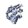
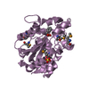
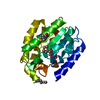
| Structure viewer | Molecule: MolmilJmol/JSmol |
|---|
- Downloads & links
-Download
| PDBx/mmCIF format | 2c1f.cif.gz | 61.1 KB | Display | PDBx/mmCIF format |
|---|---|---|---|---|
| PDB format | pdb2c1f.ent.gz | 43.3 KB | Display | PDB format |
| PDBx/mmJSON format | 2c1f.json.gz | Tree view | PDBx/mmJSON format | |
| Others |  Other downloads Other downloads |
-Validation report
| Arichive directory | https://data.pdbj.org/pub/pdb/validation_reports/c1/2c1fftp://data.pdbj.org/pub/pdb/validation_reports/c1/2c1f | HTTPS FTP |
|---|
-Related structure data
| Related structure data |  2vg9C  2vgdC  1f5jS S: Starting model for refinement C: citing same article ( |
|---|---|
| Similar structure data |


-Links
 PDBj
PDBj
- Assembly
| Deposited unit | 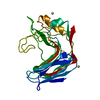
| ||||||||
|---|---|---|---|---|---|---|---|---|---|
| 1 |
| ||||||||
| Unit cell |
|
-Components
| #1: Protein | Mass: 26011.660 Da / Num. of mol.: 1 / Fragment: DOMAIN 2, RESIDUES 275-499 Source method: isolated from a genetically manipulated source Source: (gene. exp.) NEOCALLIMASTIX PATRICIARUM (fungus) / Production host:  | ||||
|---|---|---|---|---|---|
| #2: Chemical |   Mass: 112.411 Da / Num. of mol.: 2 / Source method: obtained synthetically / Formula: Cd Mass: 112.411 Da / Num. of mol.: 2 / Source method: obtained synthetically / Formula: Cd#3: Water | ChemComp-HOH / |  Mass: 18.015 Da / Num. of mol.: 173 / Source method: isolated from a natural source / Formula: H2O Mass: 18.015 Da / Num. of mol.: 173 / Source method: isolated from a natural source / Formula: H2OSequence details | CONSTRUCT CORRESPOND | |
-Experimental details
-Experiment
| Experiment | Method: X-RAY DIFFRACTION / Number of used crystals: 1 |
|---|
- Sample preparation
| Crystal | Density Matthews: 2.8 Å3/Da / Density % sol: 55 % |
|---|---|
| Crystal grow | Details: 0.1M CDCL2, 0.1M SODIUM ACETATE AND 30% V/V PEG400 |
-Data collection
| Diffraction | Mean temperature: 100 K |
|---|---|
| Diffraction source | Source: ROTATING ANODE / Type: RIGAKU / Wavelength: 1.5418 |
| Detector | Type: RIGAKU IMAGE PLATE / Detector: IMAGE PLATE / Date: Apr 14, 2004 / Details: MIRRORS |
| Radiation | Protocol: SINGLE WAVELENGTH / Monochromatic (M) / Laue (L): M / Scattering type: x-ray |
| Radiation wavelength | Wavelength: 1.5418 Å / Relative weight: 1 |
| Reflection | Resolution: 2.1→37.04 Å / Num. obs: 15357 / % possible obs: 96.8 % / Observed criterion σ(I): 0 / Redundancy: 7.49 % / Biso Wilson estimate: 26 Å2 / Rmerge(I) obs: 0.09 / Net I/σ(I): 7.37 |
| Reflection shell | Resolution: 2.1→2.21 Å / Redundancy: 2.87 % / Rmerge(I) obs: 0.36 / Mean I/σ(I) obs: 2.07 / % possible all: 82.5 |
- Processing
| Software |
| ||||||||||||||||||||||||||||||||||||||||||||||||||||||||||||||||||||||||||||||||||||||||||||||||||||||||||||||||||||||||||||||||||||||||||||||||||||||||||||||||||||||||||||||||||||||
|---|---|---|---|---|---|---|---|---|---|---|---|---|---|---|---|---|---|---|---|---|---|---|---|---|---|---|---|---|---|---|---|---|---|---|---|---|---|---|---|---|---|---|---|---|---|---|---|---|---|---|---|---|---|---|---|---|---|---|---|---|---|---|---|---|---|---|---|---|---|---|---|---|---|---|---|---|---|---|---|---|---|---|---|---|---|---|---|---|---|---|---|---|---|---|---|---|---|---|---|---|---|---|---|---|---|---|---|---|---|---|---|---|---|---|---|---|---|---|---|---|---|---|---|---|---|---|---|---|---|---|---|---|---|---|---|---|---|---|---|---|---|---|---|---|---|---|---|---|---|---|---|---|---|---|---|---|---|---|---|---|---|---|---|---|---|---|---|---|---|---|---|---|---|---|---|---|---|---|---|---|---|---|---|
| Refinement | Method to determine structure: MOLECULAR REPLACEMENT Starting model: PDB ENTRY 1F5J Resolution: 2.1→83.05 Å / Cor.coef. Fo:Fc: 0.958 / Cor.coef. Fo:Fc free: 0.935 / SU B: 8.951 / SU ML: 0.106 / TLS residual ADP flag: LIKELY RESIDUAL / Cross valid method: THROUGHOUT / σ(F): 0 / ESU R: 0.186 / ESU R Free: 0.159 / Stereochemistry target values: MAXIMUM LIKELIHOOD / Details: HYDROGENS HAVE BEEN ADDED IN THE RIDING POSITIONS.
| ||||||||||||||||||||||||||||||||||||||||||||||||||||||||||||||||||||||||||||||||||||||||||||||||||||||||||||||||||||||||||||||||||||||||||||||||||||||||||||||||||||||||||||||||||||||
| Solvent computation | Ion probe radii: 0.8 Å / VDW probe radii: 1.2 Å / Solvent model: MASK BULK SOLVENT | ||||||||||||||||||||||||||||||||||||||||||||||||||||||||||||||||||||||||||||||||||||||||||||||||||||||||||||||||||||||||||||||||||||||||||||||||||||||||||||||||||||||||||||||||||||||
| Displacement parameters | Biso mean: 17.2 Å2 | ||||||||||||||||||||||||||||||||||||||||||||||||||||||||||||||||||||||||||||||||||||||||||||||||||||||||||||||||||||||||||||||||||||||||||||||||||||||||||||||||||||||||||||||||||||||
| Refinement step | Cycle: LAST / Resolution: 2.1→83.05 Å
| ||||||||||||||||||||||||||||||||||||||||||||||||||||||||||||||||||||||||||||||||||||||||||||||||||||||||||||||||||||||||||||||||||||||||||||||||||||||||||||||||||||||||||||||||||||||
| Refine LS restraints |
|