 Movie
Movie Controller


+ Open data

- Basic information
| Entry | Database: PDB / ID: 2bzv | ||||||
|---|---|---|---|---|---|---|---|
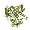
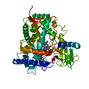
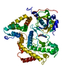
| Title | Human Enteric Adenovirus Serotype 41 Short Fiber Head (pH8) | ||||||
 Components Components | FIBER PROTEIN 2 | ||||||
 Keywords Keywords | VIRAL PROTEIN / ADENOVIRUS / FIBER / RECEPTOR / AD41 / FIBER PROTEIN | ||||||
| Function / homology |  Function and homology information Function and homology informationadhesion receptor-mediated virion attachment to host cell / viral capsid / cell adhesion / symbiont entry into host cell / host cell nucleus Similarity search - Function | ||||||
| Biological species |  HUMAN ADENOVIRUS TYPE 41 HUMAN ADENOVIRUS TYPE 41 | ||||||
| Method |  X-RAY DIFFRACTION / SYNCHROTRON / MOLECULAR REPLACEMENT / Resolution: 1.15 Å X-RAY DIFFRACTION / SYNCHROTRON / MOLECULAR REPLACEMENT / Resolution: 1.15 Å | ||||||
 Authors Authors | Seiradake, E. / Cusack, S. | ||||||
 Citation Citation | Journal: J.Virol. / Year: 2005 Title: Crystal Structure of Enteric Adenovirus Serotype 41 Short Fiber Head. Authors: Seiradake, E. / Cusack, S. | ||||||
| History |
|
- Structure visualization
| Structure viewer | Molecule: MolmilJmol/JSmol |
|---|
- Downloads & links
-Download
| PDBx/mmCIF format | 2bzv.cif.gz | 88.9 KB | Display | PDBx/mmCIF format |
|---|---|---|---|---|
| PDB format | pdb2bzv.ent.gz | 67.1 KB | Display | PDB format |
| PDBx/mmJSON format | 2bzv.json.gz | Tree view | PDBx/mmJSON format | |
| Others |  Other downloads Other downloads |
-Validation report
| Arichive directory | https://data.pdbj.org/pub/pdb/validation_reports/bz/2bzvftp://data.pdbj.org/pub/pdb/validation_reports/bz/2bzv | HTTPS FTP |
|---|
-Related structure data
| Related structure data |  2bzuSC S: Starting model for refinement C: citing same article ( |
|---|---|
| Similar structure data |


-Links
 PDBj
PDBj
- Assembly
| Deposited unit | 
| |||||||||
|---|---|---|---|---|---|---|---|---|---|---|
| 1 | 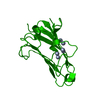
| |||||||||
| Unit cell |
| |||||||||
| Components on special symmetry positions |
|
-Components
| #1: Protein | Mass: 20148.709 Da / Num. of mol.: 1 / Fragment: RECEPTOR-BINDING DOMAIN, RESIDUES 215-387 Source method: isolated from a genetically manipulated source Source: (gene. exp.) HUMAN ADENOVIRUS TYPE 41 / Plasmid: PPROEX-HTB / Production host:  | ||
|---|---|---|---|
| #2: Water | ChemComp-HOH /  Mass: 18.015 Da / Num. of mol.: 333 / Source method: isolated from a natural source / Formula: H2O Mass: 18.015 Da / Num. of mol.: 333 / Source method: isolated from a natural source / Formula: H2O | ||
| Compound details | RECOGNIZES| Sequence details | C-TERMINAL FRAGMENT (RESIDUES 215-387) WITH C-TERMINAL HIS- TAG | |
-Experimental details
-Experiment
| Experiment | Method: X-RAY DIFFRACTION / Number of used crystals: 1 |
|---|
- Sample preparation
| Crystal | Density Matthews: 2.3 Å3/Da / Density % sol: 47 % |
|---|---|
| Crystal grow | pH: 8 Details: 20% PEG3350, 0.25 M SODIUM DIHYDROGEN PHOSPHATE MONOBASIC, pH 8.00 |
-Data collection
| Diffraction | Mean temperature: 100 K |
|---|---|
| Diffraction source | Source: SYNCHROTRON / Site: ESRF  / Beamline: ID14-2 / Wavelength: 0.933 / Beamline: ID14-2 / Wavelength: 0.933 |
| Detector | Type: ADSC CCD / Detector: CCD / Details: TOROIDAL MIRROR |
| Radiation | Monochromator: DIAMOND (111), GE(220) / Protocol: SINGLE WAVELENGTH / Monochromatic (M) / Laue (L): M / Scattering type: x-ray |
| Radiation wavelength | Wavelength: 0.933 Å / Relative weight: 1 |
| Reflection | Resolution: 1.15→100 Å / Num. obs: 351024 / % possible obs: 97 % / Observed criterion σ(I): 1 / Redundancy: 5.4 % / Rmerge(I) obs: 0.08 / Net I/σ(I): 12 |
| Reflection shell | Resolution: 1.15→1.19 Å / Redundancy: 5.1 % / Rmerge(I) obs: 0.33 / Mean I/σ(I) obs: 4 / % possible all: 92 |
- Processing
| Software |
| ||||||||||||||||||||||||||||||||||||||||||||||||||||||||||||||||||||||||||||||||||||||||||||||||||||||||||||||||||||||||||||||||||||||||||||||||||||||||||||||||||||||||||||||||||||||
|---|---|---|---|---|---|---|---|---|---|---|---|---|---|---|---|---|---|---|---|---|---|---|---|---|---|---|---|---|---|---|---|---|---|---|---|---|---|---|---|---|---|---|---|---|---|---|---|---|---|---|---|---|---|---|---|---|---|---|---|---|---|---|---|---|---|---|---|---|---|---|---|---|---|---|---|---|---|---|---|---|---|---|---|---|---|---|---|---|---|---|---|---|---|---|---|---|---|---|---|---|---|---|---|---|---|---|---|---|---|---|---|---|---|---|---|---|---|---|---|---|---|---|---|---|---|---|---|---|---|---|---|---|---|---|---|---|---|---|---|---|---|---|---|---|---|---|---|---|---|---|---|---|---|---|---|---|---|---|---|---|---|---|---|---|---|---|---|---|---|---|---|---|---|---|---|---|---|---|---|---|---|---|---|
| Refinement | Method to determine structure: MOLECULAR REPLACEMENT Starting model: PDB ENTRY 2BZU Resolution: 1.15→30 Å / Cor.coef. Fo:Fc: 0.982 / Cor.coef. Fo:Fc free: 0.981 / SU B: 0.692 / SU ML: 0.015 / Cross valid method: THROUGHOUT / ESU R: 0.027 / ESU R Free: 0.025 / Stereochemistry target values: MAXIMUM LIKELIHOOD / Details: HYDROGENS HAVE BEEN ADDED IN THE RIDING POSITIONS.
| ||||||||||||||||||||||||||||||||||||||||||||||||||||||||||||||||||||||||||||||||||||||||||||||||||||||||||||||||||||||||||||||||||||||||||||||||||||||||||||||||||||||||||||||||||||||
| Solvent computation | Ion probe radii: 0.8 Å / Shrinkage radii: 0.8 Å / VDW probe radii: 1.2 Å / Solvent model: MASK | ||||||||||||||||||||||||||||||||||||||||||||||||||||||||||||||||||||||||||||||||||||||||||||||||||||||||||||||||||||||||||||||||||||||||||||||||||||||||||||||||||||||||||||||||||||||
| Displacement parameters | Biso mean: 13.32 Å2 | ||||||||||||||||||||||||||||||||||||||||||||||||||||||||||||||||||||||||||||||||||||||||||||||||||||||||||||||||||||||||||||||||||||||||||||||||||||||||||||||||||||||||||||||||||||||
| Refinement step | Cycle: LAST / Resolution: 1.15→30 Å
| ||||||||||||||||||||||||||||||||||||||||||||||||||||||||||||||||||||||||||||||||||||||||||||||||||||||||||||||||||||||||||||||||||||||||||||||||||||||||||||||||||||||||||||||||||||||
| Refine LS restraints |
|