 Movie
Movie Controller

[English] 日本語
 Yorodumi
- PDB-2byl: Structure of ornithine aminotransferase triple mutant Y85I Y55A G320F -
+ Open data

- Basic information
| Entry | Database: PDB / ID: 2byl | ||||||
|---|---|---|---|---|---|---|---|
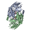
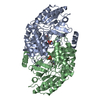
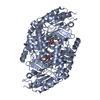
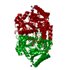
| Title | Structure of ornithine aminotransferase triple mutant Y85I Y55A G320F | ||||||
 Components Components | ORNITHINE AMINOTRANSFERASE | ||||||
 Keywords Keywords | TRANSFERASE / DISEASE MUTATION / MITOCHONDRION / TRANSIT PEPTIDE PLP-DEPENDENT ENZYME / POLYMORPHISM / PYRIDOXAL PHOSPHATE | ||||||
| Function / homology |  Function and homology information Function and homology information: / ornithine aminotransferase / L-ornithine transaminase activity / : / L-proline biosynthetic process / Glutamate and glutamine metabolism / visual perception / pyridoxal phosphate binding / mitochondrial matrix / mitochondrion ...: / ornithine aminotransferase / L-ornithine transaminase activity / : / L-proline biosynthetic process / Glutamate and glutamine metabolism / visual perception / pyridoxal phosphate binding / mitochondrial matrix / mitochondrion / nucleoplasm / identical protein binding / cytoplasm Similarity search - Function | ||||||
| Biological species |  HOMO SAPIENS (human) HOMO SAPIENS (human) | ||||||
| Method |  X-RAY DIFFRACTION / SYNCHROTRON / MOLECULAR REPLACEMENT / Resolution: 2.15 Å X-RAY DIFFRACTION / SYNCHROTRON / MOLECULAR REPLACEMENT / Resolution: 2.15 Å | ||||||
 Authors Authors | Markova, M. / Peneff, C. / Hewlins, M.J.E. / Schirmer, T. / John, R.A. | ||||||
 Citation Citation | Journal: J.Biol.Chem. / Year: 2005 Title: Determinants of Substrate Specificity in Omega-Aminotransferases. Authors: Markova, M. / Peneff, C. / Hewlins, M.J.E. / Schirmer, T. / John, R.A. | ||||||
| History |
| ||||||
| Remark 650 | HELIX DETERMINATION METHOD: AUTHOR PROVIDED. | ||||||
| Remark 700 | SHEET DETERMINATION METHOD: AUTHOR PROVIDED. |
- Structure visualization
| Structure viewer | Molecule: MolmilJmol/JSmol |
|---|
- Downloads & links
-Download
| PDBx/mmCIF format | 2byl.cif.gz | 249.7 KB | Display | PDBx/mmCIF format |
|---|---|---|---|---|
| PDB format | pdb2byl.ent.gz | 201.5 KB | Display | PDB format |
| PDBx/mmJSON format | 2byl.json.gz | Tree view | PDBx/mmJSON format | |
| Others |  Other downloads Other downloads |
-Validation report
| Arichive directory | https://data.pdbj.org/pub/pdb/validation_reports/by/2bylftp://data.pdbj.org/pub/pdb/validation_reports/by/2byl | HTTPS FTP |
|---|
-Related structure data
| Related structure data |  2byjC  1oatS S: Starting model for refinement C: citing same article ( |
|---|---|
| Similar structure data |


-Links
 PDBj
PDBj- Assembly
| Deposited unit | 
| ||||||||||||
|---|---|---|---|---|---|---|---|---|---|---|---|---|---|
| 1 | 
| ||||||||||||
| 2 | 
| ||||||||||||
| Unit cell |
| ||||||||||||
| Components on special symmetry positions |
| ||||||||||||
| Noncrystallographic symmetry (NCS) | NCS oper:
|
-Components
| #1: Protein | Mass: 48541.676 Da / Num. of mol.: 3 / Mutation: YES Source method: isolated from a genetically manipulated source Source: (gene. exp.) HOMO SAPIENS (human) / Production host:  #2: Chemical |   Mass: 247.142 Da / Num. of mol.: 3 / Source method: obtained synthetically / Formula: C8H10NO6P Mass: 247.142 Da / Num. of mol.: 3 / Source method: obtained synthetically / Formula: C8H10NO6P#3: Water | ChemComp-HOH / |  Mass: 18.015 Da / Num. of mol.: 580 / Source method: isolated from a natural source / Formula: H2O Mass: 18.015 Da / Num. of mol.: 580 / Source method: isolated from a natural source / Formula: H2OCompound details | ENGINEERED RESIDUE IN CHAIN A, TYR 85 TO ILE ENGINEERED RESIDUE IN CHAIN A, TYR 55 TO ALA ...ENGINEERED | |
|---|
-Experimental details
-Experiment
| Experiment | Method: X-RAY DIFFRACTION / Number of used crystals: 1 |
|---|
- Sample preparation
| Crystal | Density Matthews: 2.78 Å3/Da / Density % sol: 47 % |
|---|---|
| Crystal grow | pH: 7.9 Details: 6-10%PEG6000, 120-160MM NACL, 50MM TRICINE PH7.9, 1MM DTT, pH 7.90 |
-Data collection
| Diffraction | Mean temperature: 100 K |
|---|---|
| Diffraction source | Source: SYNCHROTRON / Site: SLS  / Beamline: X06SA / Wavelength: 0.977 / Beamline: X06SA / Wavelength: 0.977 |
| Detector | Type: MARRESEARCH / Detector: CCD / Date: Oct 26, 2002 |
| Radiation | Protocol: SINGLE WAVELENGTH / Monochromatic (M) / Laue (L): M / Scattering type: x-ray |
| Radiation wavelength | Wavelength: 0.977 Å / Relative weight: 1 |
| Reflection | Resolution: 2→39.22 Å / Num. obs: 78515 / % possible obs: 97.8 % / Observed criterion σ(I): 0 / Redundancy: 5.86 % / Rmerge(I) obs: 0.09 / Net I/σ(I): 5.5 |
| Reflection shell | Resolution: 2.15→2.27 Å / Redundancy: 5.17 % / Rmerge(I) obs: 0.3 / Mean I/σ(I) obs: 2.41 / % possible all: 91.4 |
- Processing
| Software |
| ||||||||||||||||||||||||||||||||||||||||||||||||||||||||||||||||||||||||||||||||||||||||||||||||||||||||||||||||||||||||||||||||||||||||||||||||||||||||||||||||||||||||||||||||||||||
|---|---|---|---|---|---|---|---|---|---|---|---|---|---|---|---|---|---|---|---|---|---|---|---|---|---|---|---|---|---|---|---|---|---|---|---|---|---|---|---|---|---|---|---|---|---|---|---|---|---|---|---|---|---|---|---|---|---|---|---|---|---|---|---|---|---|---|---|---|---|---|---|---|---|---|---|---|---|---|---|---|---|---|---|---|---|---|---|---|---|---|---|---|---|---|---|---|---|---|---|---|---|---|---|---|---|---|---|---|---|---|---|---|---|---|---|---|---|---|---|---|---|---|---|---|---|---|---|---|---|---|---|---|---|---|---|---|---|---|---|---|---|---|---|---|---|---|---|---|---|---|---|---|---|---|---|---|---|---|---|---|---|---|---|---|---|---|---|---|---|---|---|---|---|---|---|---|---|---|---|---|---|---|---|
| Refinement | Method to determine structure: MOLECULAR REPLACEMENT Starting model: PDB ENTRY 1OAT Resolution: 2.15→30 Å / Cor.coef. Fo:Fc: 0.961 / Cor.coef. Fo:Fc free: 0.944 / SU B: 7.915 / SU ML: 0.109 / Cross valid method: THROUGHOUT / ESU R: 0.191 / ESU R Free: 0.155 / Stereochemistry target values: MAXIMUM LIKELIHOOD / Details: HYDROGENS HAVE BEEN ADDED IN THE RIDING POSITIONS.
| ||||||||||||||||||||||||||||||||||||||||||||||||||||||||||||||||||||||||||||||||||||||||||||||||||||||||||||||||||||||||||||||||||||||||||||||||||||||||||||||||||||||||||||||||||||||
| Solvent computation | Ion probe radii: 0.8 Å / Shrinkage radii: 0.8 Å / VDW probe radii: 1.2 Å / Solvent model: BABINET MODEL WITH MASK | ||||||||||||||||||||||||||||||||||||||||||||||||||||||||||||||||||||||||||||||||||||||||||||||||||||||||||||||||||||||||||||||||||||||||||||||||||||||||||||||||||||||||||||||||||||||
| Displacement parameters | Biso mean: 26.9 Å2
| ||||||||||||||||||||||||||||||||||||||||||||||||||||||||||||||||||||||||||||||||||||||||||||||||||||||||||||||||||||||||||||||||||||||||||||||||||||||||||||||||||||||||||||||||||||||
| Refinement step | Cycle: LAST / Resolution: 2.15→30 Å
| ||||||||||||||||||||||||||||||||||||||||||||||||||||||||||||||||||||||||||||||||||||||||||||||||||||||||||||||||||||||||||||||||||||||||||||||||||||||||||||||||||||||||||||||||||||||
| Refine LS restraints |
|