 Movie
Movie Controller

[English] 日本語
 Yorodumi
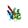
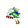
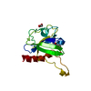
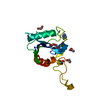
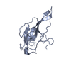
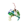
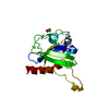
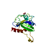
- PDB-2bxx: Crystal structure of the N-terminal domain of IBV coronavirus nuc... -
+ Open data

- Basic information
| Entry | Database: PDB / ID: 2bxx | ||||||
|---|---|---|---|---|---|---|---|
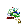
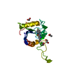
| Title | Crystal structure of the N-terminal domain of IBV coronavirus nucleocapsid. Native crystal form | ||||||
 Components Components | NUCLEOCAPSID PROTEIN | ||||||
 Keywords Keywords | NUCLEOCAPSID PROTEIN / PHOSPHORYLATION / RNA-BINDING / VIRAL NUCLEOPROTEIN | ||||||
| Function / homology |  Function and homology information Function and homology informationviral nucleocapsid / host cell endoplasmic reticulum-Golgi intermediate compartment / host cell Golgi apparatus / ribonucleoprotein complex / DNA-templated transcription / RNA binding / identical protein binding Similarity search - Function | ||||||
| Biological species |  AVIAN INFECTIOUS BRONCHITIS VIRUS AVIAN INFECTIOUS BRONCHITIS VIRUS | ||||||
| Method |  X-RAY DIFFRACTION / MOLECULAR REPLACEMENT / Resolution: 1.85 Å X-RAY DIFFRACTION / MOLECULAR REPLACEMENT / Resolution: 1.85 Å | ||||||
 Authors Authors | Fan, H. / Ooi, A. / Liu, D.-X. / Lescar, J. | ||||||
 Citation Citation | Journal: Structure / Year: 2005 Title: The Nucleocapsid Protein of Coronavirus Infectious Bronchitis Virus: Crystal Structure of its N-Terminal Domain and Multimerization Properties. Authors: Fan, H. / Ooi, A. / Tan, Y.W. / Wang, S. / Fang, S. / Liu, D.-X. / Lescar, J. | ||||||
| History |
|
- Structure visualization
| Structure viewer | Molecule: MolmilJmol/JSmol |
|---|
- Downloads & links
-Download
| PDBx/mmCIF format | 2bxx.cif.gz | 67.9 KB | Display | PDBx/mmCIF format |
|---|---|---|---|---|
| PDB format | pdb2bxx.ent.gz | 50 KB | Display | PDB format |
| PDBx/mmJSON format | 2bxx.json.gz | Tree view | PDBx/mmJSON format | |
| Others |  Other downloads Other downloads |
-Validation report
| Arichive directory | https://data.pdbj.org/pub/pdb/validation_reports/bx/2bxxftp://data.pdbj.org/pub/pdb/validation_reports/bx/2bxx | HTTPS FTP |
|---|
-Related structure data
| Related structure data |  2btlSC S: Starting model for refinement C: citing same article ( |
|---|---|
| Similar structure data |
-Links
 PDBj
PDBj- Assembly
| Deposited unit | 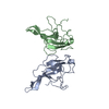
| ||||||||
|---|---|---|---|---|---|---|---|---|---|
| 1 | 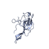
| ||||||||
| 2 | 
| ||||||||
| Unit cell |
| ||||||||
| Noncrystallographic symmetry (NCS) | NCS oper: (Code: given Matrix: (0.376, -0.927, 0.003), Vector: |
-Components
| #1: Protein | Mass: 15014.549 Da / Num. of mol.: 2 / Fragment: RNA BINDING DOMAIN RESIDUES 29-160 / Mutation: YES Source method: isolated from a genetically manipulated source Source: (gene. exp.) AVIAN INFECTIOUS BRONCHITIS VIRUS / Strain: BEAUDETTE / Production host:  #2: Water | ChemComp-HOH / |  Mass: 18.015 Da / Num. of mol.: 187 / Source method: isolated from a natural source / Formula: H2O Mass: 18.015 Da / Num. of mol.: 187 / Source method: isolated from a natural source / Formula: H2OCompound details | ENGINEERED | Sequence details | ENGINEERED | |
|---|
-Experimental details
-Experiment
| Experiment | Method: X-RAY DIFFRACTION |
|---|
- Sample preparation
| Crystal | Density Matthews: 2.02 Å3/Da / Density % sol: 38.62 % |
|---|
-Data collection
| Diffraction | Mean temperature: 100 K |
|---|---|
| Diffraction source | Source: ROTATING ANODE / Type: RIGAKU MICROMAX-007 / Wavelength: 1.5418 |
| Detector | Detector: IMAGE PLATE / Date: Dec 10, 2004 |
| Radiation | Protocol: SINGLE WAVELENGTH / Monochromatic (M) / Laue (L): M / Scattering type: x-ray |
| Radiation wavelength | Wavelength: 1.5418 Å / Relative weight: 1 |
| Reflection | Resolution: 1.85→20 Å / Num. obs: 20031 / % possible obs: 92.4 % / Observed criterion σ(I): 0 / Redundancy: 3.8 % / Rmerge(I) obs: 0.06 / Net I/σ(I): 7.4 |
| Reflection shell | Resolution: 1.85→1.95 Å / Redundancy: 3.7 % / Rmerge(I) obs: 0.62 / Mean I/σ(I) obs: 1.1 / % possible all: 88.8 |
- Processing
| Software | Name: CNS / Version: 1.1 / Classification: refinement | ||||||||||||||||||||||||||||||||||||||||||||||||||||||||||||
|---|---|---|---|---|---|---|---|---|---|---|---|---|---|---|---|---|---|---|---|---|---|---|---|---|---|---|---|---|---|---|---|---|---|---|---|---|---|---|---|---|---|---|---|---|---|---|---|---|---|---|---|---|---|---|---|---|---|---|---|---|---|
| Refinement | Method to determine structure: MOLECULAR REPLACEMENT Starting model: PDB ENTRY 2BTL Resolution: 1.85→19.92 Å / Cross valid method: THROUGHOUT / Stereochemistry target values: MAXIMUM LIKEIHOOD / Details: HYDROGENS HAVE BEEN ADDED IN THE RIDING POSITIONS
| ||||||||||||||||||||||||||||||||||||||||||||||||||||||||||||
| Solvent computation | Solvent model: BABINET MODEL WITH MASK | ||||||||||||||||||||||||||||||||||||||||||||||||||||||||||||
| Displacement parameters | Biso mean: 34.284 Å2
| ||||||||||||||||||||||||||||||||||||||||||||||||||||||||||||
| Refinement step | Cycle: LAST / Resolution: 1.85→19.92 Å
| ||||||||||||||||||||||||||||||||||||||||||||||||||||||||||||
| Refine LS restraints |
|
