[English] 日本語
Yorodumi
- PDB-2btl: Crystal structure of the N-terminal domain of IBV coronavirus nuc... -

+
Open data


ID or keywords:

Loading...

-
Basic information

Entry
Database: PDB / ID: 2btl
TitleCrystal structure of the N-terminal domain of IBV coronavirus nucleocapsid
ComponentsINFECTIOUS BRONCHITIS VIRUS NUCLEOCAPSID PROTEIN
KeywordsNUCLEOCAPSID PROTEIN / PHOSPHORYLATION / RNA-BINDING / VIRAL NUCLEOPROTEIN
Function / homology
Function and homology information


host cell endoplasmic reticulum-Golgi intermediate compartment / host cell Golgi apparatus / viral nucleocapsid / ribonucleoprotein complex / RNA binding / identical protein binding
Similarity search - Function
Nucleocapsid protein, gammacoronavirus / Nucleocapsid (N) protein, C-terminal domain, coronavirus / Nucleocapsid (N) protein, N-terminal domain, coronavirus / Coronavirus nucleocapsid (CoV N) protein N-terminal (NTD) domain profile. / Coronavirus nucleocapsid (CoV N) protein C-terminal (CTD) domain profile. / Nucleocapsid protein, coronavirus / Nucleocapsid protein, C-terminal / Nucleocapsid protein, N-terminal / Coronavirus nucleocapsid
Similarity search - Domain/homology
Biological speciesAVIAN INFECTIOUS BRONCHITIS VIRUS
MethodX-RAY DIFFRACTION / SYNCHROTRON / MAD / Resolution: 1.95 Å
AuthorsFan, H. / Ooi, A. / Liu, D.-X. / Lescar, J.
CitationJournal: Structure / Year: 2005
Title: The Nucleocapsid Protein of Coronavirus Infectious Bronchitis Virus: Crystal Structure of its N-Terminal Domain and Multimerization Properties.
Authors: Fan, H. / Ooi, A. / Tan, Y.W. / Wang, S. / Fang, S. / Liu, D.-X. / Lescar, J.
History
DepositionJun 3, 2005Deposition site: PDBE / Processing site: PDBE
Revision 1.0Dec 14, 2005Provider: repository / Type: Initial release
Revision 1.1May 8, 2011Group: Version format compliance
Revision 1.2Jul 13, 2011Group: Version format compliance
Revision 1.3May 8, 2024Group: Data collection / Database references ...Data collection / Database references / Other / Refinement description
Category: chem_comp_atom / chem_comp_bond ...chem_comp_atom / chem_comp_bond / database_2 / pdbx_database_status / struct_ncs_dom_lim
Item: _database_2.pdbx_DOI / _database_2.pdbx_database_accession ..._database_2.pdbx_DOI / _database_2.pdbx_database_accession / _pdbx_database_status.status_code_sf / _struct_ncs_dom_lim.beg_auth_comp_id / _struct_ncs_dom_lim.beg_label_asym_id / _struct_ncs_dom_lim.beg_label_comp_id / _struct_ncs_dom_lim.beg_label_seq_id / _struct_ncs_dom_lim.end_auth_comp_id / _struct_ncs_dom_lim.end_label_asym_id / _struct_ncs_dom_lim.end_label_comp_id / _struct_ncs_dom_lim.end_label_seq_id

-
Structure visualization

Structure viewerMolecule:
MolmilJmol/JSmol

Downloads & links

-
Assembly

Deposited unit
A: INFECTIOUS BRONCHITIS VIRUS NUCLEOCAPSID PROTEIN
B: INFECTIOUS BRONCHITIS VIRUS NUCLEOCAPSID PROTEIN


Theoretical massNumber of molelcules
Total (without water)30,1652
Polymers30,1652
Non-polymers00
Water3,279182
1
A: INFECTIOUS BRONCHITIS VIRUS NUCLEOCAPSID PROTEIN


Theoretical massNumber of molelcules
Total (without water)15,0831
Polymers15,0831
Non-polymers00
Water181
TypeNameSymmetry operationNumber
identity operation1_555x,y,z1
MethodPQS
2
B: INFECTIOUS BRONCHITIS VIRUS NUCLEOCAPSID PROTEIN


Theoretical massNumber of molelcules
Total (without water)15,0831
Polymers15,0831
Non-polymers00
Water181
TypeNameSymmetry operationNumber
identity operation1_555x,y,z1
MethodPQS
Unit cell
Length a, b, c (Å)34.770, 35.370, 55.950
Angle α, β, γ (deg.)100.51, 95.48, 110.16
Int Tables number1
Space group name H-MP1
Noncrystallographic symmetry (NCS)NCS domain:
IDEns-IDDetails
11A
21B

NCS domain segments:

Ens-ID: 1 / Refine code: 4

Dom-IDComponent-IDBeg auth comp-IDBeg label comp-IDEnd auth comp-IDEnd label comp-IDAuth asym-IDLabel asym-IDAuth seq-IDLabel seq-ID
11SERSERALAALAAA29 - 333 - 7
21SERSERALAALABB29 - 333 - 7
12LYSLYSPROPROAA43 - 4817 - 22
22LYSLYSPROPROBB43 - 4817 - 22
13SERSERGLNGLNAA65 - 7439 - 48
23SERSERGLNGLNBB65 - 7439 - 48
14PROPROTRPTRPAA86 - 9160 - 65
24PROPROTRPTRPBB86 - 9160 - 65

NCS oper: (Code: given
Matrix: (0.37591, -0.92665, 0.00305), (-0.92629, -0.37585, -0.02685), (0.02603, 0.00726, -0.99963)
Vector: 1.56722, -3.17674, 16.0061)

-
Components

#1: Protein INFECTIOUS BRONCHITIS VIRUS NUCLEOCAPSID PROTEIN / N STRUCTURAL PROTEIN / NC


Mass: 15082.688 Da / Num. of mol.: 2 / Fragment: RNA BINDING DOMAIN, RESIDUES 29-160 / Mutation: YES
Source method: isolated from a genetically manipulated source
Source: (gene. exp.) AVIAN INFECTIOUS BRONCHITIS VIRUS / Strain: BEAUDETTE / Production host: ESCHERICHIA COLI (E. coli) / References: UniProt: P69596
#2: Water ChemComp-HOH / water


Mass: 18.015 Da / Num. of mol.: 182 / Source method: isolated from a natural source / Formula: H2O
Compound details
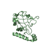
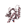
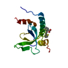
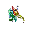
FUNCTION: MAJOR STRUCTURAL COMPONENT OF VIRIONS THAT FORMS A HELICAL NUCLEOCAPSID WHEN ASSOCIATED ...FUNCTION: MAJOR STRUCTURAL COMPONENT OF VIRIONS THAT FORMS A HELICAL NUCLEOCAPSID WHEN ASSOCIATED WITH GENOMIC RNA ENGINEERED RESIDUE IN CHAIN A, ILE 62 TO MET ENGINEERED RESIDUE IN CHAIN A, LYS 85 TO CYS ENGINEERED RESIDUE IN CHAIN A, LEU 104 TO MET ENGINEERED RESIDUE IN CHAIN A, VAL 116 TO MET ENGINEERED RESIDUE IN CHAIN B, ILE 62 TO MET ENGINEERED RESIDUE IN CHAIN B, LYS 85 TO CYS ENGINEERED RESIDUE IN CHAIN B, LEU 104 TO MET ENGINEERED RESIDUE IN CHAIN B, VAL 116 TO MET
Sequence detailsOUR CONSTRUCT HAS ENGINEERED MUTATIONS COMPARED TO THE DEPOSITED SEQUENCE IN GENBANK

-
Experimental details

-
Experiment

ExperimentMethod: X-RAY DIFFRACTION / Number of used crystals: 1

-
Sample preparation

CrystalDensity Matthews: 1.93 Å3/Da / Density % sol: 40.6 %

-
Data collection

DiffractionMean temperature: 100 K
Diffraction sourceSource: SYNCHROTRON / Site: Photon Factory / Beamline: AR-NW12A / Wavelength: 0.97943
DetectorType: ADSC CCD / Detector: CCD / Date: Dec 20, 2004 / Details: MIRRORS
RadiationProtocol: MAD / Monochromatic (M) / Laue (L): M / Scattering type: x-ray
Radiation wavelengthWavelength: 0.97943 Å / Relative weight: 1
ReflectionResolution: 1.95→20 Å / Num. obs: 76265 / % possible obs: 96.6 % / Observed criterion σ(I): 0 / Redundancy: 3.8 % / Rmerge(I) obs: 0.05 / Net I/σ(I): 8.6
Reflection shellResolution: 1.95→2 Å / Redundancy: 3.7 % / Rmerge(I) obs: 0.12 / Mean I/σ(I) obs: 3.7 / % possible all: 95

-
Processing

Software
NameVersionClassification
REFMAC5.2.0005refinement
MOSFLMdata reduction
SCALAdata scaling
SOLVEphasing
RefinementMethod to determine structure: MAD / Resolution: 1.95→20 Å / Cor.coef. Fo:Fc: 0.935 / Cor.coef. Fo:Fc free: 0.914 / SU B: 6.576 / SU ML: 0.187 / Cross valid method: THROUGHOUT / ESU R: 0.266 / ESU R Free: 0.21 / Stereochemistry target values: MAXIMUM LIKELIHOOD / Details: HYDROGENS HAVE BEEN ADDED IN THE RIDING POSITIONS.
RfactorNum. reflection% reflectionSelection details
Rfree0.276 881 5.2 %RANDOM
Rwork0.227 ---
obs0.23 16077 96.1 %-
Solvent computationIon probe radii: 0.8 Å / Shrinkage radii: 0.8 Å / VDW probe radii: 1.2 Å / Solvent model: BABINET MODEL WITH MASK
Displacement parametersBiso mean: 30.63 Å2
Baniso -1Baniso -2Baniso -3
1--2.29 Å20 Å20.71 Å2
2--2.02 Å22.84 Å2
3---1.45 Å2
Refinement stepCycle: LAST / Resolution: 1.95→20 Å
ProteinNucleic acidLigandSolventTotal
Num. atoms2128 0 0 182 2310
Refine LS restraints
Refine-IDTypeDev idealDev ideal targetNumber
X-RAY DIFFRACTIONr_bond_refined_d0.0080.0222206
X-RAY DIFFRACTIONr_bond_other_d
X-RAY DIFFRACTIONr_angle_refined_deg1.1441.9222992
X-RAY DIFFRACTIONr_angle_other_deg
X-RAY DIFFRACTIONr_dihedral_angle_1_deg6.2665266
X-RAY DIFFRACTIONr_dihedral_angle_2_deg31.92723.509114
X-RAY DIFFRACTIONr_dihedral_angle_3_deg14.24515324
X-RAY DIFFRACTIONr_dihedral_angle_4_deg11.9981516
X-RAY DIFFRACTIONr_chiral_restr0.0770.2266
X-RAY DIFFRACTIONr_gen_planes_refined0.0040.021816
X-RAY DIFFRACTIONr_gen_planes_other
X-RAY DIFFRACTIONr_nbd_refined0.2030.2821
X-RAY DIFFRACTIONr_nbd_other
X-RAY DIFFRACTIONr_nbtor_refined0.3010.21454
X-RAY DIFFRACTIONr_nbtor_other
X-RAY DIFFRACTIONr_xyhbond_nbd_refined0.160.2156
X-RAY DIFFRACTIONr_xyhbond_nbd_other
X-RAY DIFFRACTIONr_metal_ion_refined
X-RAY DIFFRACTIONr_metal_ion_other
X-RAY DIFFRACTIONr_symmetry_vdw_refined0.1830.280
X-RAY DIFFRACTIONr_symmetry_vdw_other
X-RAY DIFFRACTIONr_symmetry_hbond_refined0.1750.216
X-RAY DIFFRACTIONr_symmetry_hbond_other
X-RAY DIFFRACTIONr_symmetry_metal_ion_refined
X-RAY DIFFRACTIONr_symmetry_metal_ion_other
X-RAY DIFFRACTIONr_mcbond_it0.6911.51363
X-RAY DIFFRACTIONr_mcbond_other
X-RAY DIFFRACTIONr_mcangle_it1.21422127
X-RAY DIFFRACTIONr_mcangle_other
X-RAY DIFFRACTIONr_scbond_it1.29331006
X-RAY DIFFRACTIONr_scbond_other
X-RAY DIFFRACTIONr_scangle_it1.9124.5865
X-RAY DIFFRACTIONr_scangle_other
X-RAY DIFFRACTIONr_long_range_B_refined
X-RAY DIFFRACTIONr_long_range_B_other
X-RAY DIFFRACTIONr_rigid_bond_restr
X-RAY DIFFRACTIONr_sphericity_free
X-RAY DIFFRACTIONr_sphericity_bonded
Refine LS restraints NCS

Dom-ID: 1 / Auth asym-ID: A / Ens-ID: 1 / Number: 231 / Refine-ID: X-RAY DIFFRACTION

TypeRms dev position (Å)Weight position
medium positional0.210.5
medium thermal1.522
LS refinement shellResolution: 1.95→2 Å / Total num. of bins used: 20 /
RfactorNum. reflection
Rfree0.393 62
Rwork0.282 1158

+
About Yorodumi

-
News

-
Feb 9, 2022. New format data for meta-information of EMDB entries

New format data for meta-information of EMDB entries

  • Version 3 of the EMDB header file is now the official format.
  • The previous official version 1.9 will be removed from the archive.

Related info.:EMDB header

External links:wwPDB to switch to version 3 of the EMDB data model

-
Aug 12, 2020. Covid-19 info

Covid-19 info

URL: https://pdbjlvh1.pdbj.org/emnavi/covid19.php

New page: Covid-19 featured information page in EM Navigator.

Related info.:Covid-19 info / Mar 5, 2020. Novel coronavirus structure data

+
Mar 5, 2020. Novel coronavirus structure data

Novel coronavirus structure data

Related info.:Yorodumi Speices / Aug 12, 2020. Covid-19 info

External links:COVID-19 featured content - PDBj / Molecule of the Month (242):Coronavirus Proteases

+
Jan 31, 2019. EMDB accession codes are about to change! (news from PDBe EMDB page)

EMDB accession codes are about to change! (news from PDBe EMDB page)

  • The allocation of 4 digits for EMDB accession codes will soon come to an end. Whilst these codes will remain in use, new EMDB accession codes will include an additional digit and will expand incrementally as the available range of codes is exhausted. The current 4-digit format prefixed with “EMD-” (i.e. EMD-XXXX) will advance to a 5-digit format (i.e. EMD-XXXXX), and so on. It is currently estimated that the 4-digit codes will be depleted around Spring 2019, at which point the 5-digit format will come into force.
  • The EM Navigator/Yorodumi systems omit the EMD- prefix.

Related info.:Q: What is EMD? / ID/Accession-code notation in Yorodumi/EM Navigator

External links:EMDB Accession Codes are Changing Soon! / Contact to PDBj

+
Jul 12, 2017. Major update of PDB

Major update of PDB

  • wwPDB released updated PDB data conforming to the new PDBx/mmCIF dictionary.
  • This is a major update changing the version number from 4 to 5, and with Remediation, in which all the entries are updated.
  • In this update, many items about electron microscopy experimental information are reorganized (e.g. em_software).
  • Now, EM Navigator and Yorodumi are based on the updated data.

External links:wwPDB Remediation / Enriched Model Files Conforming to OneDep Data Standards Now Available in the PDB FTP Archive

-
Yorodumi

Thousand views of thousand structures

  • Yorodumi is a browser for structure data from EMDB, PDB, SASBDB, etc.
  • This page is also the successor to EM Navigator detail page, and also detail information page/front-end page for Omokage search.
  • The word "yorodu" (or yorozu) is an old Japanese word meaning "ten thousand". "mi" (miru) is to see.

Related info.:EMDB / PDB / SASBDB / Comparison of 3 databanks / Yorodumi Search / Aug 31, 2016. New EM Navigator & Yorodumi / Yorodumi Papers / Jmol/JSmol / Function and homology information / Changes in new EM Navigator and Yorodumi

Read more