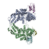
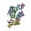
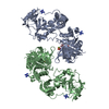
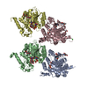
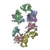
- PDB-2bw3: Three-dimensional structure of the Hermes DNA transposase -
+
Open data
ID or keywords:
Loading...
-
Basic information
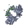
Entry
Database: PDB / ID: 2bw3
Title
Three-dimensional structure of the Hermes DNA transposase
Components
(TRANSPOSASE) x 2
Keywords
DNA RECOMBINATION / TRANSPOSITION
Function / homology
Function and homology information
protein dimerization activity / regulation of transcription by RNA polymerase II / DNA-templated transcription / DNA binding / zinc ion binding / identical protein binding / nucleus Similarity search - Function
: / Zinc finger, BED domain-containing / : / Hermes trasposase, DNA-binding domain / Hermes transposase DNA-binding domain / HAT, C-terminal dimerisation domain / hAT family C-terminal dimerisation region / Zinc finger, BED-type / Zinc finger BED-type profile. / BED zinc finger ...: / Zinc finger, BED domain-containing / : / Hermes trasposase, DNA-binding domain / Hermes transposase DNA-binding domain / HAT, C-terminal dimerisation domain / hAT family C-terminal dimerisation region / Zinc finger, BED-type / Zinc finger BED-type profile. / BED zinc finger / Arc Repressor Mutant, subunit A / Ribonuclease H-like superfamily / Orthogonal Bundle / Mainly Alpha Similarity search - Domain/homology
Mass: 18.015 Da / Num. of mol.: 416 / Source method: isolated from a natural source / Formula: H2O
Compound details
ENGINEERED RESIDUE IN CHAIN A, SER 163 TO GLY ENGINEERED RESIDUE IN CHAIN A, LEU 233 TO SER ...ENGINEERED RESIDUE IN CHAIN A, SER 163 TO GLY ENGINEERED RESIDUE IN CHAIN A, LEU 233 TO SER ENGINEERED RESIDUE IN CHAIN A, VAL 286 TO MET
Has protein modification
Y
-
Experimental details
-
Experiment
Experiment
Method: X-RAY DIFFRACTION / Number of used crystals: 1
-
Sample preparation
Crystal
Density Matthews: 2.57 Å3/Da / Density % sol: 49.2 %
In the structure databanks used in Yorodumi, some data are registered as the other names, "COVID-19 virus" and "2019-nCoV". Here are the details of the virus and the list of structure data.
Jan 31, 2019. EMDB accession codes are about to change! (news from PDBe EMDB page)
EMDB accession codes are about to change! (news from PDBe EMDB page)
The allocation of 4 digits for EMDB accession codes will soon come to an end. Whilst these codes will remain in use, new EMDB accession codes will include an additional digit and will expand incrementally as the available range of codes is exhausted. The current 4-digit format prefixed with “EMD-” (i.e. EMD-XXXX) will advance to a 5-digit format (i.e. EMD-XXXXX), and so on. It is currently estimated that the 4-digit codes will be depleted around Spring 2019, at which point the 5-digit format will come into force.
The EM Navigator/Yorodumi systems omit the EMD- prefix.
Related info.:Q: What is EMD? / ID/Accession-code notation in Yorodumi/EM Navigator
Yorodumi is a browser for structure data from EMDB, PDB, SASBDB, etc.
This page is also the successor to EM Navigator detail page, and also detail information page/front-end page for Omokage search.
The word "yorodu" (or yorozu) is an old Japanese word meaning "ten thousand". "mi" (miru) is to see.
Related info.:EMDB / PDB / SASBDB / Comparison of 3 databanks / Yorodumi Search / Aug 31, 2016. New EM Navigator & Yorodumi / Yorodumi Papers / Jmol/JSmol / Function and homology information / Changes in new EM Navigator and Yorodumi
 Movie
Movie Controller


 Open data
 Basic information
 Components
 Keywords
 Function and homology information
 MUSCA DOMESTICA (house fly)
 X-RAY DIFFRACTION /
  Authors
 Citation
 Structure visualization
 Downloads & links
 Other downloads
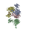

 PDBj
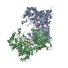
PDBj Assembly
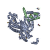

 Mass: 18.015 Da / Num. of mol.: 416 / Source method: isolated from a natural source / Formula: H2O
 Sample preparation
 / Beamline: 22-ID / Wavelength: 0.9686
  Processing