 Movie
Movie Controller

[English] 日本語
 Yorodumi
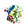
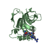
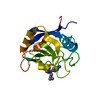
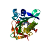
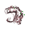
- PDB-2bsw: Crystal structure of a glyphosate-N-acetyltransferase obtained by... -
+ Open data

- Basic information
| Entry | Database: PDB / ID: 2bsw | ||||||
|---|---|---|---|---|---|---|---|
| Title | Crystal structure of a glyphosate-N-acetyltransferase obtained by DNA shuffling. | ||||||
 Components Components | GLYPHOSATE N-ACETYLTRANSFERASE | ||||||
 Keywords Keywords | TRANSFERASE / GNAT SUPERFAMILY / GLYPHOSATE N-ACETYLTRANSFERASE | ||||||
| Function / homology |  Function and homology information Function and homology informationacyltransferase activity, transferring groups other than amino-acyl groups Similarity search - Function | ||||||
| Biological species | SYNTHETIC CONSTRUCT (others) | ||||||
| Method |  X-RAY DIFFRACTION / SYNCHROTRON / SAD / Resolution: 1.63 Å X-RAY DIFFRACTION / SYNCHROTRON / SAD / Resolution: 1.63 Å | ||||||
 Authors Authors | Keenan, R.J. / Siehl, D.L. / Gorton, R. / Castle, L.A. | ||||||
 Citation Citation | Journal: Proc.Natl.Acad.Sci.USA / Year: 2005 Title: DNA Shuffling as a Tool for Protein Crystallization. Authors: Keenan, R.J. / Siehl, D.L. / Gorton, R. / Castle, L.A. | ||||||
| History |
|
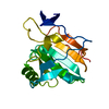
- Structure visualization
| Structure viewer | Molecule: MolmilJmol/JSmol |
|---|
- Downloads & links
-Download
| PDBx/mmCIF format | 2bsw.cif.gz | 50.1 KB | Display | PDBx/mmCIF format |
|---|---|---|---|---|
| PDB format | pdb2bsw.ent.gz | 35.3 KB | Display | PDB format |
| PDBx/mmJSON format | 2bsw.json.gz | Tree view | PDBx/mmJSON format | |
| Others |  Other downloads Other downloads |
-Validation report
| Arichive directory | https://data.pdbj.org/pub/pdb/validation_reports/bs/2bswftp://data.pdbj.org/pub/pdb/validation_reports/bs/2bsw | HTTPS FTP |
|---|
-Related structure data
| Similar structure data |
|---|


-Links
 PDBj
PDBj

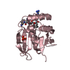
- Assembly
| Deposited unit | 
| |||||||||
|---|---|---|---|---|---|---|---|---|---|---|
| 1 |
| |||||||||
| Unit cell |
| |||||||||
| Components on special symmetry positions |
|
-Components
| #1: Protein | Mass: 16812.572 Da / Num. of mol.: 1 Source method: isolated from a genetically manipulated source Source: (gene. exp.) SYNTHETIC CONSTRUCT (others) Description: THE ENZYME IS A SHUFFLED VARIANT DERIVED FROM GENES DISCOVERED IN B. LICHENIFORMIS. Plasmid: PQE80 / Production host:  | ||||
|---|---|---|---|---|---|
| #2: Chemical | ChemComp-CAO / 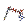  Mass: 783.534 Da / Num. of mol.: 1 / Source method: obtained synthetically / Formula: C21H36N7O17P3S Mass: 783.534 Da / Num. of mol.: 1 / Source method: obtained synthetically / Formula: C21H36N7O17P3S | ||||
| #3: Chemical | ChemComp-SO4 /   Mass: 96.063 Da / Num. of mol.: 1 / Source method: obtained synthetically / Formula: SO4 Mass: 96.063 Da / Num. of mol.: 1 / Source method: obtained synthetically / Formula: SO4 | ||||
| #4: Chemical |   Mass: 92.094 Da / Num. of mol.: 2 / Source method: obtained synthetically / Formula: C3H8O3 Mass: 92.094 Da / Num. of mol.: 2 / Source method: obtained synthetically / Formula: C3H8O3#5: Water | ChemComp-HOH / |  Mass: 18.015 Da / Num. of mol.: 161 / Source method: isolated from a natural source / Formula: H2O Mass: 18.015 Da / Num. of mol.: 161 / Source method: isolated from a natural source / Formula: H2OHas protein modification | Y | |
-Experimental details
-Experiment
| Experiment | Method: X-RAY DIFFRACTION / Number of used crystals: 1 |
|---|
- Sample preparation
| Crystal | Density Matthews: 2.3 Å3/Da / Density % sol: 46.57 % |
|---|---|
| Crystal grow | pH: 4.6 Details: 100 MM SODIUM ACETATE, PH 4.6 250 MM AMMONIUM SULFATE 25% PEG 4000 |
-Data collection
| Diffraction | Mean temperature: 100 K |
|---|---|
| Diffraction source | Source: SYNCHROTRON / Site: ALS  / Beamline: 5.0.2 / Wavelength: 0.97942 / Beamline: 5.0.2 / Wavelength: 0.97942 |
| Detector | Type: ADSC CCD / Detector: CCD / Date: Dec 12, 2002 |
| Radiation | Protocol: SINGLE WAVELENGTH / Monochromatic (M) / Laue (L): M / Scattering type: x-ray |
| Radiation wavelength | Wavelength: 0.97942 Å / Relative weight: 1 |
| Reflection | Resolution: 1.63→50 Å / Num. obs: 18939 / % possible obs: 97.4 % / Observed criterion σ(I): 0 / Redundancy: 2.94 % / Rmerge(I) obs: 0.07 / Net I/σ(I): 20.2 |
| Reflection shell | Resolution: 1.63→1.69 Å / Rmerge(I) obs: 0.16 / Mean I/σ(I) obs: 8 / % possible all: 98.1 |
- Processing
| Software |
| ||||||||||||||||||||||||||||||||||||||||||||||||||||||||||||||||||||||||||||||||||||||||||||||||||||||||||||||||||||||||||||||||||||||||||||||||||||||||||||||||||||||||||||||||||||||
|---|---|---|---|---|---|---|---|---|---|---|---|---|---|---|---|---|---|---|---|---|---|---|---|---|---|---|---|---|---|---|---|---|---|---|---|---|---|---|---|---|---|---|---|---|---|---|---|---|---|---|---|---|---|---|---|---|---|---|---|---|---|---|---|---|---|---|---|---|---|---|---|---|---|---|---|---|---|---|---|---|---|---|---|---|---|---|---|---|---|---|---|---|---|---|---|---|---|---|---|---|---|---|---|---|---|---|---|---|---|---|---|---|---|---|---|---|---|---|---|---|---|---|---|---|---|---|---|---|---|---|---|---|---|---|---|---|---|---|---|---|---|---|---|---|---|---|---|---|---|---|---|---|---|---|---|---|---|---|---|---|---|---|---|---|---|---|---|---|---|---|---|---|---|---|---|---|---|---|---|---|---|---|---|
| Refinement | Method to determine structure: SAD / Resolution: 1.63→45.17 Å / Cor.coef. Fo:Fc: 0.96 / Cor.coef. Fo:Fc free: 0.949 / SU B: 1.774 / SU ML: 0.063 / Cross valid method: THROUGHOUT / ESU R: 0.102 / ESU R Free: 0.1 / Stereochemistry target values: MAXIMUM LIKELIHOOD Details: HYDROGENS HAVE BEEN ADDED IN THE RIDING POSITIONS. THE N-TERMINAL SELENOMETHIONINE, MSE A1, IS DISORDERED IN ELECTRON DENSITY MAPS.
| ||||||||||||||||||||||||||||||||||||||||||||||||||||||||||||||||||||||||||||||||||||||||||||||||||||||||||||||||||||||||||||||||||||||||||||||||||||||||||||||||||||||||||||||||||||||
| Solvent computation | Ion probe radii: 0.8 Å / Shrinkage radii: 0.8 Å / VDW probe radii: 1.2 Å / Solvent model: MASK | ||||||||||||||||||||||||||||||||||||||||||||||||||||||||||||||||||||||||||||||||||||||||||||||||||||||||||||||||||||||||||||||||||||||||||||||||||||||||||||||||||||||||||||||||||||||
| Displacement parameters | Biso mean: 18.65 Å2
| ||||||||||||||||||||||||||||||||||||||||||||||||||||||||||||||||||||||||||||||||||||||||||||||||||||||||||||||||||||||||||||||||||||||||||||||||||||||||||||||||||||||||||||||||||||||
| Refinement step | Cycle: LAST / Resolution: 1.63→45.17 Å
| ||||||||||||||||||||||||||||||||||||||||||||||||||||||||||||||||||||||||||||||||||||||||||||||||||||||||||||||||||||||||||||||||||||||||||||||||||||||||||||||||||||||||||||||||||||||
| Refine LS restraints |
|