 Movie
Movie Controller

[English] 日本語
 Yorodumi
- PDB-2bse: Structure of Lactococcal Bacteriophage p2 Receptor Binding Protei... -
+ Open data

- Basic information
| Entry | Database: PDB / ID: 2bse | ||||||
|---|---|---|---|---|---|---|---|
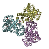
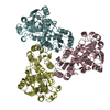
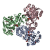
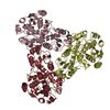
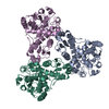
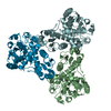
| Title | Structure of Lactococcal Bacteriophage p2 Receptor Binding Protein in complex with a llama VHH domain | ||||||
 Components Components |
| ||||||
 Keywords Keywords | RECEPTOR / LACTOCOCCUS LACTIS / PHAGE / RECEPTOR BINDING PROTEIN / LLAMA ANTIBODY / VHH | ||||||
| Function / homology |  Function and homology information Function and homology informationvirus tail, baseplate / entry receptor-mediated virion attachment to host cell / cell adhesion / symbiont entry into host cell / virion attachment to host cell Similarity search - Function | ||||||
| Biological species |  LACTOCOCCUS VIRUS P2 LACTOCOCCUS VIRUS P2 | ||||||
| Method |  X-RAY DIFFRACTION / SYNCHROTRON / MOLECULAR REPLACEMENT / Resolution: 2.7 Å X-RAY DIFFRACTION / SYNCHROTRON / MOLECULAR REPLACEMENT / Resolution: 2.7 Å | ||||||
 Authors Authors | Spinelli, S. / Desmyter, A. / Verrips, C.T. / de Haard, H.J.W. / Moineau, S. / Cambillau, C. | ||||||
 Citation Citation | Journal: Nat.Struct.Mol.Biol. / Year: 2006 Title: Lactococcal Bacteriophage P2 Receptor Binding Protein Structure Suggests a Common Ancestor Gene with Bacterial and Mammalian Viruses. Authors: Spinelli, S. / Desmyter, A. / Verrips, C.T. / de Haard, H.J.W. / Moineau, S. / Cambillau, C. | ||||||
| History |
| ||||||
| Remark 700 | SHEET THE SHEET STRUCTURE OF THIS MOLECULE IS BIFURCATED. IN ORDER TO REPRESENT THIS FEATURE IN ... SHEET THE SHEET STRUCTURE OF THIS MOLECULE IS BIFURCATED. IN ORDER TO REPRESENT THIS FEATURE IN THE SHEET RECORDS BELOW, TWO SHEETS ARE DEFINED. |
- Structure visualization
| Structure viewer | Molecule: MolmilJmol/JSmol |
|---|
- Downloads & links
-Download
| PDBx/mmCIF format | 2bse.cif.gz | 142.9 KB | Display | PDBx/mmCIF format |
|---|---|---|---|---|
| PDB format | pdb2bse.ent.gz | 110.3 KB | Display | PDB format |
| PDBx/mmJSON format | 2bse.json.gz | Tree view | PDBx/mmJSON format | |
| Others |  Other downloads Other downloads |
-Validation report
| Arichive directory | https://data.pdbj.org/pub/pdb/validation_reports/bs/2bseftp://data.pdbj.org/pub/pdb/validation_reports/bs/2bse | HTTPS FTP |
|---|
-Related structure data
| Related structure data |  2bsdSC S: Starting model for refinement C: citing same article ( |
|---|---|
| Similar structure data |
-Links
 PDBj
PDBj


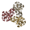
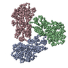
- Assembly
| Deposited unit | 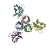
| ||||||||
|---|---|---|---|---|---|---|---|---|---|
| 1 |
| ||||||||
| Unit cell |
|
-Components
| #1: Protein | Mass: 28668.057 Da / Num. of mol.: 3 Source method: isolated from a genetically manipulated source Source: (gene. exp.) LACTOCOCCUS VIRUS P2 / Production host:  #2: Antibody | Mass: 13499.944 Da / Num. of mol.: 3 / Fragment: VHH RECOGNITION DOMAIN, RESIDUES 1-123 Source method: isolated from a genetically manipulated source Source: (gene. exp.)  #3: Water | ChemComp-HOH / |  Mass: 18.015 Da / Num. of mol.: 1 / Source method: isolated from a natural source / Formula: H2O Mass: 18.015 Da / Num. of mol.: 1 / Source method: isolated from a natural source / Formula: H2OHas protein modification | Y | |
|---|
-Experimental details
-Experiment
| Experiment | Method: X-RAY DIFFRACTION / Number of used crystals: 1 |
|---|
- Sample preparation
| Crystal | Density Matthews: 2.95 Å3/Da |
|---|---|
| Crystal grow | pH: 6.5 / Details: pH 6.50 |
-Data collection
| Diffraction | Mean temperature: 100 K |
|---|---|
| Diffraction source | Source: SYNCHROTRON / Site: ESRF  / Beamline: ID14-4 / Wavelength: 0.9794 / Beamline: ID14-4 / Wavelength: 0.9794 |
| Detector | Type: ADSC CCD / Detector: CCD |
| Radiation | Protocol: SINGLE WAVELENGTH / Monochromatic (M) / Laue (L): M / Scattering type: x-ray |
| Radiation wavelength | Wavelength: 0.9794 Å / Relative weight: 1 |
| Reflection | Resolution: 2.7→15 Å / Num. obs: 23055 / % possible obs: 99 % / Observed criterion σ(I): 1 / Redundancy: 2.3 % / Rmerge(I) obs: 0.1 / Net I/σ(I): 5.6 |
| Reflection shell | Resolution: 2.7→2.77 Å / Rmerge(I) obs: 0.3 / Mean I/σ(I) obs: 2 |
- Processing
| Software |
| ||||||||||||||||||||||||||||||||||||||||||||||||||||||||||||||||||||||||||||||||||||||||||||||||||||||||||||||||||||||||||||||||||||||||||||||||||||||||||||||||||||||||||||||||||||||
|---|---|---|---|---|---|---|---|---|---|---|---|---|---|---|---|---|---|---|---|---|---|---|---|---|---|---|---|---|---|---|---|---|---|---|---|---|---|---|---|---|---|---|---|---|---|---|---|---|---|---|---|---|---|---|---|---|---|---|---|---|---|---|---|---|---|---|---|---|---|---|---|---|---|---|---|---|---|---|---|---|---|---|---|---|---|---|---|---|---|---|---|---|---|---|---|---|---|---|---|---|---|---|---|---|---|---|---|---|---|---|---|---|---|---|---|---|---|---|---|---|---|---|---|---|---|---|---|---|---|---|---|---|---|---|---|---|---|---|---|---|---|---|---|---|---|---|---|---|---|---|---|---|---|---|---|---|---|---|---|---|---|---|---|---|---|---|---|---|---|---|---|---|---|---|---|---|---|---|---|---|---|---|---|
| Refinement | Method to determine structure: MOLECULAR REPLACEMENT Starting model: PDB ENTRY 2BSD Resolution: 2.7→15 Å / Cor.coef. Fo:Fc: 0.907 / Cor.coef. Fo:Fc free: 0.884 / SU B: 16.719 / SU ML: 0.336 / TLS residual ADP flag: LIKELY RESIDUAL / Cross valid method: THROUGHOUT / ESU R: 1.192 / ESU R Free: 0.388 / Stereochemistry target values: MAXIMUM LIKELIHOOD / Details: HYDROGENS HAVE BEEN ADDED IN THE RIDING POSITIONS.
| ||||||||||||||||||||||||||||||||||||||||||||||||||||||||||||||||||||||||||||||||||||||||||||||||||||||||||||||||||||||||||||||||||||||||||||||||||||||||||||||||||||||||||||||||||||||
| Solvent computation | Ion probe radii: 0.8 Å / Shrinkage radii: 0.8 Å / VDW probe radii: 1.4 Å / Solvent model: BABINET MODEL WITH MASK | ||||||||||||||||||||||||||||||||||||||||||||||||||||||||||||||||||||||||||||||||||||||||||||||||||||||||||||||||||||||||||||||||||||||||||||||||||||||||||||||||||||||||||||||||||||||
| Displacement parameters | Biso mean: 23.72 Å2
| ||||||||||||||||||||||||||||||||||||||||||||||||||||||||||||||||||||||||||||||||||||||||||||||||||||||||||||||||||||||||||||||||||||||||||||||||||||||||||||||||||||||||||||||||||||||
| Refinement step | Cycle: LAST / Resolution: 2.7→15 Å
| ||||||||||||||||||||||||||||||||||||||||||||||||||||||||||||||||||||||||||||||||||||||||||||||||||||||||||||||||||||||||||||||||||||||||||||||||||||||||||||||||||||||||||||||||||||||
| Refine LS restraints |
|