 Movie
Movie Controller


+ Open data

- Basic information
| Entry | Database: PDB / ID: 2bs2 | ||||||||||||
|---|---|---|---|---|---|---|---|---|---|---|---|---|---|
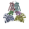
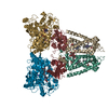
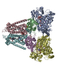
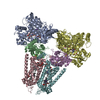
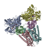
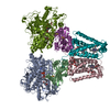
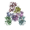
| Title | QUINOL:FUMARATE REDUCTASE FROM WOLINELLA SUCCINOGENES | ||||||||||||
 Components Components | (QUINOL-FUMARATE REDUCTASE ...) x 3 | ||||||||||||
 Keywords Keywords | OXIDOREDUCTASE / RESPIRATORY CHAIN / CITRIC ACID CYCLE / IRON-SULPHUR PROTEIN | ||||||||||||
| Function / homology |  Function and homology information Function and homology informationsuccinate dehydrogenase (quinone) activity / succinate dehydrogenase / anaerobic respiration / 3 iron, 4 sulfur cluster binding / tricarboxylic acid cycle / respiratory electron transport chain / electron transport chain / 2 iron, 2 sulfur cluster binding / flavin adenine dinucleotide binding / 4 iron, 4 sulfur cluster binding ...succinate dehydrogenase (quinone) activity / succinate dehydrogenase / anaerobic respiration / 3 iron, 4 sulfur cluster binding / tricarboxylic acid cycle / respiratory electron transport chain / electron transport chain / 2 iron, 2 sulfur cluster binding / flavin adenine dinucleotide binding / 4 iron, 4 sulfur cluster binding / electron transfer activity / metal ion binding / plasma membrane Similarity search - Function | ||||||||||||
| Biological species |  WOLINELLA SUCCINOGENES (bacteria) WOLINELLA SUCCINOGENES (bacteria) | ||||||||||||
| Method |  X-RAY DIFFRACTION / SYNCHROTRON / FOURIER SYNTHESIS / Resolution: 1.78 Å X-RAY DIFFRACTION / SYNCHROTRON / FOURIER SYNTHESIS / Resolution: 1.78 Å | ||||||||||||
 Authors Authors | Lancaster, C.R.D. | ||||||||||||
 Citation Citation | Journal: Embo J. / Year: 2006 Title: Evidence for Transmembrane Proton Transfer in a Dihaem-Containing Membrane Protein Complex. Authors: Madej, M.G. / Nasiri, H.R. / Hilgendorff, N.S. / Schwalbe, H. / Lancaster, C.R.D. #1: Journal: Biochim.Biophys.Acta / Year: 2002 Title: Wolinella Succinogenes Quinol:Fumarate Reductase -2.2 Angstrom Resolution Crystal Structure and the E-Pathway Hypothesis of Coupled Transmembrane Proton and Electron Transfer Authors: Lancaster, C.R.D. #2: Journal: Eur.J.Biochem. / Year: 2001Title: A Third Crystal Form of Wolinella Succinogenes Quinol:Fumarate Reductase Reveals Domain Closure at the Site of Fumarate Reduction Authors: Lancaster, C.R.D. / Gross, R. / Simon, J. #3: Journal: Proc.Natl.Acad.Sci.USA / Year: 2000 Title: Essential Role of Glu-C66 for Menaquinol Oxidation Indicates Transmembrane Electrochemical Potential Generation by Wolinella Succinogenes Fumarate Reductase Authors: Lancaster, C.R.D. / Gross, R. / Haas, A. / Ritter, M. / Maentele, W. / Simon, J. / Kroeger, A. #4: Journal: Nature / Year: 1999Title: Structure of Fumarate Reductase from Wolinella Succinogenes at 2.2 Angstroms Resolution Authors: Lancaster, C.R.D. / Kroeger, A. / Auer, M. / Michel, H. | ||||||||||||
| History |
|
- Structure visualization
| Structure viewer | Molecule: MolmilJmol/JSmol |
|---|
- Downloads & links
-Download
| PDBx/mmCIF format | 2bs2.cif.gz | 499.9 KB | Display | PDBx/mmCIF format |
|---|---|---|---|---|
| PDB format | pdb2bs2.ent.gz | 399.4 KB | Display | PDB format |
| PDBx/mmJSON format | 2bs2.json.gz | Tree view | PDBx/mmJSON format | |
| Others |  Other downloads Other downloads |
-Validation report
| Arichive directory | https://data.pdbj.org/pub/pdb/validation_reports/bs/2bs2ftp://data.pdbj.org/pub/pdb/validation_reports/bs/2bs2 | HTTPS FTP |
|---|
-Related structure data
| Related structure data |  1qla S: Starting model for refinement |
|---|---|
| Similar structure data |


-Links
 PDBj
PDBj


- Assembly
| Deposited unit | 
| ||||||||
|---|---|---|---|---|---|---|---|---|---|
| 1 |
| ||||||||
| Unit cell |
| ||||||||
| Noncrystallographic symmetry (NCS) | NCS oper: (Code: given Matrix: (-0.999932, -0.011239, 0.003138), Vector: |
-Components
-QUINOL-FUMARATE REDUCTASE ... , 3 types, 6 molecules ADBECF
| #1: Protein | Mass: 73424.766 Da / Num. of mol.: 2 / Source method: isolated from a natural source Details: FAD COVALENTLY BOUND TO HIS A43 BY AN 8-ALPHA-(N-EPSILON-HISTIDYL) BOND Source: (natural) WOLINELLA SUCCINOGENES (bacteria) / References: UniProt: P17412, EC: 1.3.99.1#2: Protein | Mass: 27415.727 Da / Num. of mol.: 2 / Source method: isolated from a natural source / Source: (natural) WOLINELLA SUCCINOGENES (bacteria) / References: UniProt: P17596, EC: 1.3.99.1#3: Protein | Mass: 29721.945 Da / Num. of mol.: 2 / Source method: isolated from a natural source / Source: (natural) WOLINELLA SUCCINOGENES (bacteria) / References: UniProt: P17413, EC: 1.3.99.1 |
|---|
-Sugars , 1 types, 2 molecules 
| #11: Sugar |  Type: D-saccharide / Mass: 510.615 Da / Num. of mol.: 2 / Source method: obtained synthetically / Formula: C24H46O11 / Comment: detergent*YM Type: D-saccharide / Mass: 510.615 Da / Num. of mol.: 2 / Source method: obtained synthetically / Formula: C24H46O11 / Comment: detergent*YM |
|---|
-Non-polymers , 8 types, 1006 molecules 


| #4: Chemical |  Mass: 785.550 Da / Num. of mol.: 2 / Source method: obtained synthetically / Formula: C27H33N9O15P2 / Comment: FAD*YM Mass: 785.550 Da / Num. of mol.: 2 / Source method: obtained synthetically / Formula: C27H33N9O15P2 / Comment: FAD*YM#5: Chemical |  Mass: 116.072 Da / Num. of mol.: 2 / Source method: obtained synthetically / Formula: C4H4O4 Mass: 116.072 Da / Num. of mol.: 2 / Source method: obtained synthetically / Formula: C4H4O4#6: Chemical |  Mass: 22.990 Da / Num. of mol.: 2 / Source method: obtained synthetically / Formula: Na Mass: 22.990 Da / Num. of mol.: 2 / Source method: obtained synthetically / Formula: Na#7: Chemical |  Mass: 175.820 Da / Num. of mol.: 2 / Source method: obtained synthetically / Formula: Fe2S2 Mass: 175.820 Da / Num. of mol.: 2 / Source method: obtained synthetically / Formula: Fe2S2#8: Chemical |  Mass: 295.795 Da / Num. of mol.: 2 / Source method: obtained synthetically / Formula: Fe3S4 Mass: 295.795 Da / Num. of mol.: 2 / Source method: obtained synthetically / Formula: Fe3S4#9: Chemical |  Mass: 351.640 Da / Num. of mol.: 2 / Source method: obtained synthetically / Formula: Fe4S4 Mass: 351.640 Da / Num. of mol.: 2 / Source method: obtained synthetically / Formula: Fe4S4#10: Chemical | ChemComp-HEM /  Mass: 616.487 Da / Num. of mol.: 4 / Source method: obtained synthetically / Formula: C34H32FeN4O4 Mass: 616.487 Da / Num. of mol.: 4 / Source method: obtained synthetically / Formula: C34H32FeN4O4#12: Water | ChemComp-HOH / | Mass: 18.015 Da / Num. of mol.: 990 / Source method: isolated from a natural source / Formula: H2O |
|---|
-Details
| Has protein modification | Y |
|---|
-Experimental details
-Experiment
| Experiment | Method: X-RAY DIFFRACTION / Number of used crystals: 2 |
|---|
- Sample preparation
| Crystal | Density Matthews: 3.4 Å3/Da / Density % sol: 62.4 % |
|---|---|
| Crystal grow | pH: 6 / Details: pH 6.00 |
-Data collection
| Diffraction | Mean temperature: 277 K |
|---|---|
| Diffraction source | Source: SYNCHROTRON / Site: ESRF  / Beamline: ID14-3 / Wavelength: 0.93107 / Beamline: ID14-3 / Wavelength: 0.93107 |
| Detector | Type: ADSC CCD / Detector: CCD / Date: Apr 22, 2001 |
| Radiation | Protocol: SINGLE WAVELENGTH / Monochromatic (M) / Laue (L): M / Scattering type: x-ray |
| Radiation wavelength | Wavelength: 0.93107 Å / Relative weight: 1 |
| Reflection | Resolution: 1.78→28.75 Å / Num. obs: 317968 / % possible obs: 92.3 % / Observed criterion σ(I): 0 / Redundancy: 3.8 % / Biso Wilson estimate: 23.2 Å2 / Rmerge(I) obs: 0.1 / Net I/σ(I): 16.5 |
- Processing
| Software |
| ||||||||||||||||||||||||||||||||||||||||||||||||||||||||||||
|---|---|---|---|---|---|---|---|---|---|---|---|---|---|---|---|---|---|---|---|---|---|---|---|---|---|---|---|---|---|---|---|---|---|---|---|---|---|---|---|---|---|---|---|---|---|---|---|---|---|---|---|---|---|---|---|---|---|---|---|---|---|
| Refinement | Method to determine structure: FOURIER SYNTHESIS Starting model: PDB ENTRY 1QLA 1qla Resolution: 1.78→28.7 Å / Rfactor Rfree error: 0.004 / Data cutoff high absF: 2909818.71 / Data cutoff low absF: 0 / Isotropic thermal model: RESTRAINED / Cross valid method: THROUGHOUT / σ(F): 0
| ||||||||||||||||||||||||||||||||||||||||||||||||||||||||||||
| Solvent computation | Solvent model: FLAT MODEL / Bsol: 73.9811 Å2 / ksol: 0.375084 e/Å3 | ||||||||||||||||||||||||||||||||||||||||||||||||||||||||||||
| Displacement parameters | Biso mean: 38.8 Å2
| ||||||||||||||||||||||||||||||||||||||||||||||||||||||||||||
| Refine analyze |
| ||||||||||||||||||||||||||||||||||||||||||||||||||||||||||||
| Refinement step | Cycle: LAST / Resolution: 1.78→28.7 Å
| ||||||||||||||||||||||||||||||||||||||||||||||||||||||||||||
| Refine LS restraints |
| ||||||||||||||||||||||||||||||||||||||||||||||||||||||||||||
| LS refinement shell | Resolution: 1.78→1.82 Å / Rfactor Rfree error: 0.026 / Total num. of bins used: 15
| ||||||||||||||||||||||||||||||||||||||||||||||||||||||||||||
| Xplor file |
|
