+ Open data
- Basic information
| Entry | Database: PDB / ID: 2bht | ||||||
|---|---|---|---|---|---|---|---|
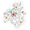
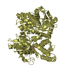
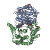
| Title | Crystal structure of O-acetylserine sulfhydrylase B | ||||||
 Components Components | CYSTEINE SYNTHASE B | ||||||
 Keywords Keywords | TRANSFERASE / CYSTEINE BIOSYNTHESIS / PLP-DEPENDENT ENZYME / PYRIDOXAL-5'-PHOSPHATE / PYRIDOXAL PHOSPHATE | ||||||
| Function / homology |  Function and homology information Function and homology informationcysteine biosynthetic process via S-sulfo-L-cysteine / L-cysteine catabolic process to pyruvate / cysteine synthase / L-cysteine desulfhydrase activity / cysteine biosynthetic process / cysteine synthase activity / cysteine biosynthetic process from serine / iron-sulfur cluster assembly / pyridoxal phosphate binding / protein homodimerization activity / cytoplasm Similarity search - Function | ||||||
| Biological species |  | ||||||
| Method |  X-RAY DIFFRACTION / X-RAY DIFFRACTION /  SYNCHROTRON / SYNCHROTRON /  MOLECULAR REPLACEMENT / Resolution: 2.1 Å MOLECULAR REPLACEMENT / Resolution: 2.1 Å | ||||||
 Authors Authors | Claus, M.T. / Zocher, G.E. / Maier, T.H.P. / Schulz, G.E. | ||||||
 Citation Citation |  Journal: Biochemistry / Year: 2005 Journal: Biochemistry / Year: 2005Title: Structure of the O-Acetylserine Sulfhydrylase Isoenzyme Cysm from Escherichia Coli Authors: Claus, M.T. / Zocher, G.E. / Maier, T.H.P. / Schulz, G.E. | ||||||
| History |
|
- Structure visualization
| Structure viewer | Molecule:  Molmil Molmil Jmol/JSmol Jmol/JSmol |
|---|
- Downloads & links
- Download
| PDBx/mmCIF format |  2bht.cif.gz 2bht.cif.gz | 229.3 KB | Display |  PDBx/mmCIF format PDBx/mmCIF format |
|---|---|---|---|---|
| PDB format |  pdb2bht.ent.gz pdb2bht.ent.gz | 186.6 KB | Display |  PDB format PDB format |
| PDBx/mmJSON format |  2bht.json.gz 2bht.json.gz | Tree view |  PDBx/mmJSON format PDBx/mmJSON format | |
| Others |  Other downloads Other downloads |
-Validation report
| Arichive directory |  https://data.pdbj.org/pub/pdb/validation_reports/bh/2bht https://data.pdbj.org/pub/pdb/validation_reports/bh/2bht ftp://data.pdbj.org/pub/pdb/validation_reports/bh/2bht ftp://data.pdbj.org/pub/pdb/validation_reports/bh/2bht | HTTPS FTP |
|---|
-Related structure data
| Related structure data |  2bhsSC S: Starting model for refinement C: citing same article ( |
|---|---|
| Similar structure data |
- Links
Links
- Assembly
| Deposited unit | 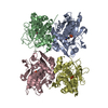
| ||||||||||||||||
|---|---|---|---|---|---|---|---|---|---|---|---|---|---|---|---|---|---|
| 1 | 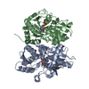
| ||||||||||||||||
| 2 | 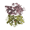
| ||||||||||||||||
| Unit cell |
| ||||||||||||||||
| Noncrystallographic symmetry (NCS) | NCS oper:
|
- Components
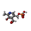
| #1: Protein | Mass: 32665.105 Da / Num. of mol.: 4 / Mutation: YES Source method: isolated from a genetically manipulated source Details: SCHIFF BASE LINK BETWEEN A41 AND PLP320 / Source: (gene. exp.)   #2: Chemical | ChemComp-PLP / #3: Water | ChemComp-HOH / | Compound details | ENGINEERED RESIDUE IN CHAIN A, GLU 57 TO ARG ENGINEERED RESIDUE IN CHAIN A, TYR 148 TO LYS ...ENGINEERED | |
|---|
-Experimental details
-Experiment
| Experiment | Method:  X-RAY DIFFRACTION / Number of used crystals: 1 X-RAY DIFFRACTION / Number of used crystals: 1 |
|---|
- Sample preparation
| Crystal | Density Matthews: 4.2 Å3/Da / Density % sol: 70 % |
|---|---|
| Crystal grow | pH: 7.6 / Details: pH 7.60 |
-Data collection
| Diffraction | Mean temperature: 100 K |
|---|---|
| Diffraction source | Source:  SYNCHROTRON / Site: SYNCHROTRON / Site:  SLS SLS  / Beamline: X06SA / Wavelength: 0.82 / Beamline: X06SA / Wavelength: 0.82 |
| Detector | Type: MARRESEARCH / Detector: CCD |
| Radiation | Protocol: SINGLE WAVELENGTH / Monochromatic (M) / Laue (L): M / Scattering type: x-ray |
| Radiation wavelength | Wavelength: 0.82 Å / Relative weight: 1 |
| Reflection | Resolution: 2.1→100 Å / Num. obs: 123872 / % possible obs: 99.8 % / Observed criterion σ(I): 2 / Redundancy: 3.9 % / Rmerge(I) obs: 0.06 / Net I/σ(I): 14.9 |
- Processing
| Software |
| |||||||||||||||||||||||||||||||||
|---|---|---|---|---|---|---|---|---|---|---|---|---|---|---|---|---|---|---|---|---|---|---|---|---|---|---|---|---|---|---|---|---|---|---|
| Refinement | Method to determine structure:  MOLECULAR REPLACEMENT MOLECULAR REPLACEMENTStarting model: PDB ENTRY 2BHS Resolution: 2.1→20 Å / Cross valid method: THROUGHOUT / σ(F): 2
| |||||||||||||||||||||||||||||||||
| Refinement step | Cycle: LAST / Resolution: 2.1→20 Å
| |||||||||||||||||||||||||||||||||
| Refine LS restraints |
|
 Movie
Movie Controller






 PDBj
PDBj