 Movie
Movie Controller

[English] 日本語
 Yorodumi
- PDB-2bat: THE STRUCTURE OF THE COMPLEX BETWEEN INFLUENZA VIRUS NEURAMINIDAS... -
+ Open data

- Basic information
| Entry | Database: PDB / ID: 2bat | |||||||||
|---|---|---|---|---|---|---|---|---|---|---|
| Title | THE STRUCTURE OF THE COMPLEX BETWEEN INFLUENZA VIRUS NEURAMINIDASE AND SIALIC ACID, THE VIRAL RECEPTOR | |||||||||
 Components Components | NEURAMINIDASE N2 | |||||||||
 Keywords Keywords | HYDROLASE(O-GLYCOSYL) | |||||||||
| Function / homology |  Function and homology information Function and homology informationexo-alpha-sialidase / exo-alpha-sialidase activity / viral budding from plasma membrane / carbohydrate metabolic process / host cell plasma membrane / virion membrane / membrane / metal ion binding Similarity search - Function | |||||||||
| Biological species |   Influenza A virus Influenza A virus | |||||||||
| Method |  X-RAY DIFFRACTION / Resolution: 2 Å X-RAY DIFFRACTION / Resolution: 2 Å | |||||||||
 Authors Authors | Varghese, J.N. / Colman, P.M. | |||||||||
 Citation Citation | Journal: Proteins / Year: 1992 Title: The structure of the complex between influenza virus neuraminidase and sialic acid, the viral receptor. Authors: Varghese, J.N. / McKimm-Breschkin, J.L. / Caldwell, J.B. / Kortt, A.A. / Colman, P.M. #1: Journal: J.Mol.Biol. / Year: 1991Title: Three-Dimensional Structure of the Neuraminidase of Influenza Virus A(Slash)Tokyo(Slash)3(Slash)67 at 2.2 Angstroms Resolution Authors: Varghese, J.N. / Colman, P.M. #2: Journal: Biochem.J. / Year: 1982Title: Amino Acid Sequence of the Pronase-Released Heads of Neuraminidase Subtype N2 from the Asian Strain A(Slash)Tokyo(Slash)3(Slash)67 of Influenza Virus Authors: Ward, C.W. / Elleman, T.C. / Azad, A.A. | |||||||||
| History |
|
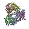
- Structure visualization
| Structure viewer | Molecule: MolmilJmol/JSmol |
|---|
- Downloads & links
-Download
| PDBx/mmCIF format | 2bat.cif.gz | 102.4 KB | Display | PDBx/mmCIF format |
|---|---|---|---|---|
| PDB format | pdb2bat.ent.gz | 75.7 KB | Display | PDB format |
| PDBx/mmJSON format | 2bat.json.gz | Tree view | PDBx/mmJSON format | |
| Others |  Other downloads Other downloads |
-Validation report
| Arichive directory | https://data.pdbj.org/pub/pdb/validation_reports/ba/2batftp://data.pdbj.org/pub/pdb/validation_reports/ba/2bat | HTTPS FTP |
|---|
-Related structure data
| Related structure data | |
|---|---|
| Similar structure data |


-Links
 PDBj
PDBj

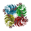
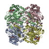
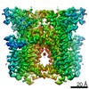
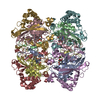
- Assembly
| Deposited unit | 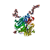
| ||||||||
|---|---|---|---|---|---|---|---|---|---|
| 1 | 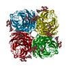
| ||||||||
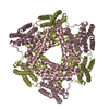
| Unit cell |
| ||||||||
| Atom site foot note | 1: RESIDUES PRO 285 AND PRO 326 ARE CIS PROLINES. 2: A NON-PROLINE CIS PEPTIDE BOND BETWEEN RESIDUES 430 AND 431 HAS BEEN POSITIVELY IDENTIFIED. PEPTIDE BOND DEVIATES SIGNIFICANTLY FROM TRANS CONFORMATION ARG 430 - LYS 431 0.445 |
-Components
-Protein , 1 types, 1 molecules A
| #1: Protein | Mass: 43140.027 Da / Num. of mol.: 1 Source method: isolated from a genetically manipulated source Source: (gene. exp.) Influenza A virus (A/Tokyo/3/1967(H2N2))Strain: A/Tokyo/3/1967(H2N2) / References: UniProt: P06820, exo-alpha-sialidase |
|---|
-Sugars , 4 types, 5 molecules 

| #2: Polysaccharide | 2-acetamido-2-deoxy-4-O-sulfo-alpha-D-galactopyranose-(1-4)-2-acetamido-2-deoxy-beta-D- ...2-acetamido-2-deoxy-4-O-sulfo-alpha-D-galactopyranose-(1-4)-2-acetamido-2-deoxy-beta-D-glucopyranose-(1-4)-alpha-D-mannopyranose-(1-3)-beta-D-mannopyranose-(1-4)-2-acetamido-2-deoxy-beta-D-glucopyranose-(1-4)-[beta-L-fucopyranose-(1-6)]2-acetamido-2-deoxy-beta-D-glucopyranose Source method: isolated from a genetically manipulated source | ||
|---|---|---|---|
| #3: Polysaccharide | alpha-D-mannopyranose-(1-2)-alpha-D-mannopyranose-(1-3)-[alpha-D-mannopyranose-(1-6)]beta-D- ...alpha-D-mannopyranose-(1-2)-alpha-D-mannopyranose-(1-3)-[alpha-D-mannopyranose-(1-6)]beta-D-mannopyranose-(1-4)-2-acetamido-2-deoxy-beta-D-glucopyranose-(1-4)-2-acetamido-2-deoxy-beta-D-glucopyranose Source method: isolated from a genetically manipulated source | ||
| #4: Sugar |  Type: D-saccharide, beta linking / Mass: 221.208 Da / Num. of mol.: 2 Type: D-saccharide, beta linking / Mass: 221.208 Da / Num. of mol.: 2Source method: isolated from a genetically manipulated source Formula: C8H15NO6 #5: Sugar | ChemComp-SIA / |  Type: D-saccharide, alpha linking / Mass: 309.270 Da / Num. of mol.: 1 Type: D-saccharide, alpha linking / Mass: 309.270 Da / Num. of mol.: 1Source method: isolated from a genetically manipulated source Formula: C11H19NO9 |
-Non-polymers , 2 types, 117 molecules 

| #6: Chemical | ChemComp-CA /  Mass: 40.078 Da / Num. of mol.: 1 / Source method: obtained synthetically / Formula: Ca Mass: 40.078 Da / Num. of mol.: 1 / Source method: obtained synthetically / Formula: Ca |
|---|---|
| #7: Water | ChemComp-HOH / Mass: 18.015 Da / Num. of mol.: 116 / Source method: isolated from a natural source / Formula: H2O |
-Details
| Has protein modification | Y |
|---|---|
| Sequence details | SEQUENCE ADVISORY NOTICE: DIFFERENCE BETWEEN SWISS-PROT AND PDB SEQUENCE. SWISS-PROT ENTRY NAME: ...SEQUENCE ADVISORY NOTICE: DIFFERENCE |
-Experimental details
-Experiment
| Experiment | Method: X-RAY DIFFRACTION |
|---|
- Sample preparation
| Crystal | Density Matthews: 5.39 Å3/Da / Density % sol: 77.19 % | |||||||||||||||||||||||||
|---|---|---|---|---|---|---|---|---|---|---|---|---|---|---|---|---|---|---|---|---|---|---|---|---|---|---|
| Crystal grow | *PLUS pH: 6.6 / Method: vapor diffusionDetails: McKimm-Breschkin, J.L. (1991) J. Virol. Methods., 32, 121. | |||||||||||||||||||||||||
| Components of the solutions | *PLUS
|
- Processing
| Software |
| ||||||||||||||||||||||||||||||||||||||||||||||||||||||||||||
|---|---|---|---|---|---|---|---|---|---|---|---|---|---|---|---|---|---|---|---|---|---|---|---|---|---|---|---|---|---|---|---|---|---|---|---|---|---|---|---|---|---|---|---|---|---|---|---|---|---|---|---|---|---|---|---|---|---|---|---|---|---|
| Refinement | Resolution: 2→6 Å / Rfactor Rwork: 0.21 / Rfactor obs: 0.21 / σ(F): 2 | ||||||||||||||||||||||||||||||||||||||||||||||||||||||||||||
| Refinement step | Cycle: LAST / Resolution: 2→6 Å
| ||||||||||||||||||||||||||||||||||||||||||||||||||||||||||||
| Refine LS restraints |
| ||||||||||||||||||||||||||||||||||||||||||||||||||||||||||||
| Refinement | *PLUS Highest resolution: 2 Å / Lowest resolution: 6 Å / σ(F): 2 / Rfactor obs: 0.21 | ||||||||||||||||||||||||||||||||||||||||||||||||||||||||||||
| Solvent computation | *PLUS | ||||||||||||||||||||||||||||||||||||||||||||||||||||||||||||
| Displacement parameters | *PLUS | ||||||||||||||||||||||||||||||||||||||||||||||||||||||||||||
| Refine LS restraints | *PLUS Type: x_angle_d / Dev ideal: 3.9 |
