[日本語] English
- PDB-2b9c: Structure of tropomyosin's mid-region: bending and binding sites ... -

+
データを開く


IDまたはキーワード:

読み込み中...

-
基本情報

登録情報
データベース: PDB / ID: 2b9c
タイトルStructure of tropomyosin's mid-region: bending and binding sites for actin
要素striated-muscle alpha tropomyosin
キーワードCONTRACTILE PROTEIN / alpha-helix / coiled coil / alanine / axial stagger / radius / side-chain packing / crystal packing / temperature factor / cardiomyopathy / elongated protein
機能・相同性
機能・相同性情報


Striated Muscle Contraction / Smooth Muscle Contraction / positive regulation of heart rate by epinephrine / bleb / negative regulation of vascular associated smooth muscle cell migration / muscle filament sliding / actin filament capping / ruffle organization / positive regulation of ATP-dependent activity / ventricular cardiac muscle tissue morphogenesis ...Striated Muscle Contraction / Smooth Muscle Contraction / positive regulation of heart rate by epinephrine / bleb / negative regulation of vascular associated smooth muscle cell migration / muscle filament sliding / actin filament capping / ruffle organization / positive regulation of ATP-dependent activity / ventricular cardiac muscle tissue morphogenesis / myofibril / sarcomere organization / negative regulation of vascular associated smooth muscle cell proliferation / positive regulation of cell adhesion / cardiac muscle contraction / stress fiber / positive regulation of stress fiber assembly / cytoskeletal protein binding / negative regulation of cell migration / muscle contraction / actin filament / actin filament organization / wound healing / ruffle membrane / cellular response to reactive oxygen species / disordered domain specific binding / actin filament binding / actin cytoskeleton / actin binding / regulation of cell shape / in utero embryonic development / protein heterodimerization activity / protein homodimerization activity / protein-containing complex / identical protein binding / cytoplasm
類似検索 - 分子機能
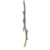
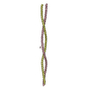
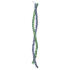
Tropomyosins signature. / Tropomyosin / Tropomyosin / Single alpha-helices involved in coiled-coils or other helix-helix interfaces - #170 / Single alpha-helices involved in coiled-coils or other helix-helix interfaces / Up-down Bundle / Mainly Alpha
類似検索 - ドメイン・相同性
Tropomyosin alpha-1 chain
類似検索 - 構成要素
生物種Rattus norvegicus (ドブネズミ)
手法X線回折 / シンクロトロン / 多波長異常分散 / 解像度: 2.3 Å
データ登録者Brown, J.H. / Zhou, Z. / Reshetnikova, L. / Robinson, H. / Yammani, R.D. / Tobacman, L.S. / Cohen, C.
引用ジャーナル: Proc.Natl.Acad.Sci.Usa / : 2005
タイトル: Structure of the mid-region of tropomyosin: Bending and binding sites for actin.
著者: Brown, J.H. / Zhou, Z. / Reshetnikova, L. / Robinson, H. / Yammani, R.D. / Tobacman, L.S. / Cohen, C.
履歴
登録2005年10月11日登録サイト: RCSB / 処理サイト: RCSB
改定 1.02006年1月3日Provider: repository / タイプ: Initial release
改定 1.12008年5月1日Group: Version format compliance
改定 1.22011年7月13日Group: Version format compliance
改定 1.32017年10月11日Group: Refinement description / カテゴリ: software
改定 1.42022年12月21日Group: Database references / カテゴリ: database_2 / struct_ref_seq_dif
Item: _database_2.pdbx_DOI / _database_2.pdbx_database_accession / _struct_ref_seq_dif.details

-
構造の表示

構造ビューア分子:
MolmilJmol/JSmol

ダウンロードとリンク

-
集合体

登録構造単位
A: striated-muscle alpha tropomyosin
B: striated-muscle alpha tropomyosin


分子量 (理論値)分子数
合計 (水以外)34,2672
ポリマ-34,2672
非ポリマー00
6,287349
1


  • 登録構造と同一
  • 登録者・ソフトウェアが定義した集合体
タイプ名称対称操作
identity operation1_555x,y,z1
Buried area6430 Å2
ΔGint-72 kcal/mol
Surface area20680 Å2
手法PISA
単位格子
Length a, b, c (Å)80.515, 80.515, 112.453
Angle α, β, γ (deg.)90.00, 90.00, 120.00
Int Tables number170
Space group name H-MP65

-
要素

#1: タンパク質 striated-muscle alpha tropomyosin


分子量: 17133.260 Da / 分子数: 2 / 由来タイプ: 組換発現 / 由来: (組換発現) Rattus norvegicus (ドブネズミ) / 発現宿主: Escherichia coli (大腸菌) / 参照: UniProt: P04692
#2: 水 ChemComp-HOH / water


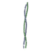
分子量: 18.015 Da / 分子数: 349 / 由来タイプ: 天然 / : H2O

-
実験情報

-
実験

実験手法: X線回折

-
試料調製

結晶マシュー密度: 3.07 Å3/Da / 溶媒含有率: 59.94 %
結晶化温度: 289 K / 手法: 蒸気拡散法, ハンギングドロップ法 / pH: 7
詳細: Native: Three microliters of protein solution [6.35mg/ml MidTm; 20mM Tris-HCl pH7.0; 100mM NaCl; 2mM 2-mercaptoethanol and 2mM NaN3] were combined with two microliters of 14% PEG2k-MME, and ...詳細: Native: Three microliters of protein solution [6.35mg/ml MidTm; 20mM Tris-HCl pH7.0; 100mM NaCl; 2mM 2-mercaptoethanol and 2mM NaN3] were combined with two microliters of 14% PEG2k-MME, and the resultant drop was equilibrated against 1ml reservoir solution [7.5% PEG2k-MME; 120mM NaCl; 24mM Tris-HCl pH7.0] by vapor diffusion at 16 C. Se-Met: Two microliters of protein solution [18mg/ml MidTm; 20mM Tris-HCl pH6.9; 100mM NaCl; 10mM mercaptoethanol and 2mM NaN3] were combined with two microliters of precipitant solution [28-32% PEG2K-MME; 50mM NaCl; 20mM Tris-HCl pH6.9 and 5mM mercaptoethanol]. The resultant mixture was equilibrated against 1ml reservoir solution [24% PEG2K-MME; 50mM NaCl; 20mM Tris-HCL pH6.9 and 5mM mercaptoethanol] by vapor diffusion at 16C. , VAPOR DIFFUSION, HANGING DROP, temperature 289K

-
データ収集

回折
ID平均測定温度 (K)Crystal-ID
11001
21001
31
放射光源
由来サイトビームラインID波長 (Å)
シンクロトロンCHESS F110.9474
シンクロトロンNSLS X29A20.9786, 0.9793, 0.9611
検出器
タイプID検出器
ADSC QUANTUM 41CCD
2CCD
放射
IDプロトコル単色(M)・ラウエ(L)散乱光タイプWavelength-ID
1SINGLE WAVELENGTHMx-ray1
2MADMx-ray2
放射波長
ID波長 (Å)相対比
10.94741
20.97861
30.97931
40.96111
Reflection
IDRmerge(I) obsΧ2D res high (Å)D res low (Å)% possible obs
184710.0931.1082.95092.6
283990.0841.0322.95091.5
382380.0931.0462.95089.3
Diffraction reflection shell
最高解像度 (Å)最低解像度 (Å)% possible obs (%)IDRmerge(I) obsChi squared
6.24509810.0671.073
4.966.2499.910.0830.996
4.334.9610010.081.175
3.944.3310010.0951.182
3.653.9499.810.1241.192
3.443.6599.810.181.171
3.273.4497.210.2511.114
3.123.2788.910.3091.069
33.1276.510.3331.035
2.9365.810.4410.962
6.245098.120.0490.958
4.966.2499.920.0780.961
4.334.9610020.0781.034
3.944.3310020.0951.021
3.653.9499.820.1281.059
3.443.6599.320.1891.146
3.273.4495.620.2711.102
3.123.2786.320.3181.049
33.1271.820.3431.022
2.9364.120.4750.944
6.245098.830.0561.104
4.966.2499.930.0841.029
4.334.9610030.0851.067
3.944.3399.930.1061.1
3.653.9499.230.1511.103
3.443.6598.530.231.038
3.273.4492.930.3470.983
3.123.2780.630.391.005
33.1264.330.4120.98
2.9358.430.590.898
反射解像度: 2.3→20 Å / Num. obs: 18253 / % possible obs: 99.4 % / Observed criterion σ(I): 2 / 冗長度: 4.7 % / Rmerge(I) obs: 0.069 / Χ2: 1.062 / Net I/σ(I): 15.9
反射 シェル解像度: 2.3→2.38 Å / 冗長度: 4.5 % / Rmerge(I) obs: 0.337 / Mean I/σ(I) obs: 4.6 / Num. measured obs: 1404 / Χ2: 1.013 / % possible all: 100

-
位相決定

位相決定手法: 多波長異常分散
Phasing set
ID
1
2
3
Phasing MAD set
Clust-IDExpt-IDSet-ID波長 (Å)F double prime refinedF prime refined
13 wavelength10.97864-5.2
13 wavelength20.97932.1-8.2
13 wavelength30.96113.8-3.4
Phasing MAD set site
IDAtom type symbolB isoFract xFract yFract zOccupancy
1Se600.5920.5380.9970.432
2Se600.7470.4150.0370.79
3Se600.6470.590.0240.645
Phasing dmFOM : 0.52 / FOM acentric: 0.52 / FOM centric: 0.56 / 反射: 7525 / Reflection acentric: 7219 / Reflection centric: 306
Phasing dm shell
解像度 (Å)FOM FOM acentricFOM centric反射Reflection acentricReflection centric
8.6-19.8030.90.90.8732529233
5.4-8.60.80.810.78103597956
4.3-5.40.730.730.71357130057
3.8-4.30.580.580.51394134351
3.2-3.80.380.380.42325225174
3-3.20.090.090.131089105435

-
解析

ソフトウェア
名称バージョン分類NB
DENZOデータ削減
SCALEPACKデータスケーリング
SOLVE2.03位相決定
RESOLVE2.03位相決定
CNS精密化
PDB_EXTRACT1.7データ抽出
精密化構造決定の手法: 多波長異常分散 / 解像度: 2.3→20 Å / σ(F): 0 / 立体化学のターゲット値: Engh & Huber
Rfactor反射数%反射Selection details
Rfree0.296 706 3.8 %random
Rwork0.256 ---
obs-17760 96.8 %-
溶媒の処理Bsol: 103.232 Å2
原子変位パラメータBiso mean: 63.133 Å2
Baniso -1Baniso -2Baniso -3
1-3.759 Å2-7.579 Å20 Å2
2--3.759 Å20 Å2
3----7.518 Å2
精密化ステップサイクル: LAST / 解像度: 2.3→20 Å
タンパク質核酸リガンド溶媒全体
原子数2301 0 0 349 2650
拘束条件
Refine-IDタイプDev ideal
X-RAY DIFFRACTIONc_bond_d0.050567
X-RAY DIFFRACTIONc_angle_deg3.86603
Xplor file
Refine-IDSerial noParam file
X-RAY DIFFRACTION1CNS_TOPPAR:protein_rep.param
X-RAY DIFFRACTION2CNS_TOPPAR:water_rep.param

+
万見について

-
お知らせ

-
2022年2月9日: EMDBエントリの付随情報ファイルのフォーマットが新しくなりました

EMDBエントリの付随情報ファイルのフォーマットが新しくなりました

  • EMDBのヘッダファイルのバージョン3が、公式のフォーマットとなりました。
  • これまでは公式だったバージョン1.9は、アーカイブから削除されます。

関連情報:EMDBヘッダ

外部リンク:wwPDBはEMDBデータモデルのバージョン3へ移行します

-
2020年8月12日: 新型コロナ情報

新型コロナ情報

URL: https://pdbjlvh1.pdbj.org/emnavi/covid19.php

新ページ: EM Navigatorに新型コロナウイルスの特設ページを開設しました。

関連情報:Covid-19情報 / 2020年3月5日: 新型コロナウイルスの構造データ

+
2020年3月5日: 新型コロナウイルスの構造データ

新型コロナウイルスの構造データ

関連情報:万見生物種 / 2020年8月12日: 新型コロナ情報

外部リンク:COVID-19特集ページ - PDBj / 今月の分子2020年2月:コロナウイルスプロテーアーゼ

+
2019年1月31日: EMDBのIDの桁数の変更

EMDBのIDの桁数の変更

  • EMDBエントリに付与されているアクセスコード(EMDB-ID)は4桁の数字(例、EMD-1234)でしたが、間もなく枯渇します。これまでの4桁のID番号は4桁のまま変更されませんが、4桁の数字を使い切った後に発行されるIDは5桁以上の数字(例、EMD-12345)になります。5桁のIDは2019年の春頃から発行される見通しです。
  • EM Navigator/万見では、接頭語「EMD-」は省略されています。

関連情報:Q: 「EMD」とは何ですか? / 万見/EM NavigatorにおけるID/アクセスコードの表記

外部リンク:EMDB Accession Codes are Changing Soon! / PDBjへお問い合わせ

+
2017年7月12日: PDB大規模アップデート

PDB大規模アップデート

  • 新バージョンのPDBx/mmCIF辞書形式に基づくデータがリリースされました。
  • 今回の更新はバージョン番号が4から5になる大規模なもので、全エントリデータの書き換えが行われる「Remediation」というアップデートに該当します。
  • このバージョンアップで、電子顕微鏡の実験手法に関する多くの項目の書式が改定されました(例:em_softwareなど)。
  • EM NavigatorとYorodumiでも、この改定に基づいた表示内容になります。

外部リンク:wwPDB Remediation / OneDepデータ基準に準拠した、より強化された内容のモデル構造ファイルが、PDBアーカイブで公開されました。

-
万見 (Yorodumi)

幾万の構造データを、幾万の視点から

  • 万見(Yorodumi)は、EMDB/PDB/SASBDBなどの構造データを閲覧するためのページです。
  • EM Navigatorの詳細ページの後継、Omokage検索のフロントエンドも兼ねています。

関連情報:EMDB / PDB / SASBDB / 3つのデータバンクの比較 / 万見検索 / 2016年8月31日: 新しいEM Navigatorと万見 / 万見文献 / Jmol/JSmol / 機能・相同性情報 / 新しいEM Navigatorと万見の変更点

他の情報も見る