[English] 日本語
Yorodumi
- PDB-2b9c: Structure of tropomyosin's mid-region: bending and binding sites ... -

+
Open data


ID or keywords:

Loading...

-
Basic information

Entry
Database: PDB / ID: 2b9c
Title
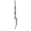
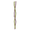
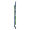
Structure of tropomyosin's mid-region: bending and binding sites for actin
Componentsstriated-muscle alpha tropomyosin
KeywordsCONTRACTILE PROTEIN / alpha-helix / coiled coil / alanine / axial stagger / radius / side-chain packing / crystal packing / temperature factor / cardiomyopathy / elongated protein
Function / homology
Function and homology information


Striated Muscle Contraction / Smooth Muscle Contraction / positive regulation of heart rate by epinephrine / bleb / negative regulation of vascular associated smooth muscle cell migration / muscle filament sliding / actin filament capping / ruffle organization / positive regulation of ATP-dependent activity / ventricular cardiac muscle tissue morphogenesis ...Striated Muscle Contraction / Smooth Muscle Contraction / positive regulation of heart rate by epinephrine / bleb / negative regulation of vascular associated smooth muscle cell migration / muscle filament sliding / actin filament capping / ruffle organization / positive regulation of ATP-dependent activity / ventricular cardiac muscle tissue morphogenesis / myofibril / sarcomere organization / negative regulation of vascular associated smooth muscle cell proliferation / positive regulation of cell adhesion / cardiac muscle contraction / stress fiber / positive regulation of stress fiber assembly / cytoskeletal protein binding / negative regulation of cell migration / muscle contraction / actin filament / actin filament organization / wound healing / ruffle membrane / cellular response to reactive oxygen species / disordered domain specific binding / actin filament binding / actin cytoskeleton / actin binding / regulation of cell shape / in utero embryonic development / protein heterodimerization activity / protein homodimerization activity / protein-containing complex / identical protein binding / cytoplasm
Similarity search - Function
Tropomyosins signature. / Tropomyosin / Tropomyosin / Single alpha-helices involved in coiled-coils or other helix-helix interfaces - #170 / Single alpha-helices involved in coiled-coils or other helix-helix interfaces / Up-down Bundle / Mainly Alpha
Similarity search - Domain/homology
Tropomyosin alpha-1 chain
Similarity search - Component
Biological speciesRattus norvegicus (Norway rat)
MethodX-RAY DIFFRACTION / SYNCHROTRON / MAD / Resolution: 2.3 Å
AuthorsBrown, J.H. / Zhou, Z. / Reshetnikova, L. / Robinson, H. / Yammani, R.D. / Tobacman, L.S. / Cohen, C.
CitationJournal: Proc.Natl.Acad.Sci.Usa / Year: 2005
Title: Structure of the mid-region of tropomyosin: Bending and binding sites for actin.
Authors: Brown, J.H. / Zhou, Z. / Reshetnikova, L. / Robinson, H. / Yammani, R.D. / Tobacman, L.S. / Cohen, C.
History
DepositionOct 11, 2005Deposition site: RCSB / Processing site: RCSB
Revision 1.0Jan 3, 2006Provider: repository / Type: Initial release
Revision 1.1May 1, 2008Group: Version format compliance
Revision 1.2Jul 13, 2011Group: Version format compliance
Revision 1.3Oct 11, 2017Group: Refinement description / Category: software
Revision 1.4Dec 21, 2022Group: Database references / Category: database_2 / struct_ref_seq_dif
Item: _database_2.pdbx_DOI / _database_2.pdbx_database_accession / _struct_ref_seq_dif.details

-
Structure visualization

Structure viewerMolecule:
MolmilJmol/JSmol

Downloads & links

-
Assembly

Deposited unit
A: striated-muscle alpha tropomyosin
B: striated-muscle alpha tropomyosin


Theoretical massNumber of molelcules
Total (without water)34,2672
Polymers34,2672
Non-polymers00
Water6,287349
1


  • Idetical with deposited unit
  • defined by author&software
TypeNameSymmetry operationNumber
identity operation1_555x,y,z1
Buried area6430 Å2
ΔGint-72 kcal/mol
Surface area20680 Å2
MethodPISA
Unit cell
Length a, b, c (Å)80.515, 80.515, 112.453
Angle α, β, γ (deg.)90.00, 90.00, 120.00
Int Tables number170
Space group name H-MP65

-
Components

#1: Protein striated-muscle alpha tropomyosin


Mass: 17133.260 Da / Num. of mol.: 2
Source method: isolated from a genetically manipulated source
Source: (gene. exp.) Rattus norvegicus (Norway rat) / Production host: Escherichia coli (E. coli) / References: UniProt: P04692
#2: Water ChemComp-HOH / water


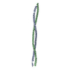
Mass: 18.015 Da / Num. of mol.: 349 / Source method: isolated from a natural source / Formula: H2O

-
Experimental details

-
Experiment

ExperimentMethod: X-RAY DIFFRACTION

-
Sample preparation

CrystalDensity Matthews: 3.07 Å3/Da / Density % sol: 59.94 %
Crystal growTemperature: 289 K / Method: vapor diffusion, hanging drop / pH: 7
Details: Native: Three microliters of protein solution [6.35mg/ml MidTm; 20mM Tris-HCl pH7.0; 100mM NaCl; 2mM 2-mercaptoethanol and 2mM NaN3] were combined with two microliters of 14% PEG2k-MME, and ...Details: Native: Three microliters of protein solution [6.35mg/ml MidTm; 20mM Tris-HCl pH7.0; 100mM NaCl; 2mM 2-mercaptoethanol and 2mM NaN3] were combined with two microliters of 14% PEG2k-MME, and the resultant drop was equilibrated against 1ml reservoir solution [7.5% PEG2k-MME; 120mM NaCl; 24mM Tris-HCl pH7.0] by vapor diffusion at 16 C. Se-Met: Two microliters of protein solution [18mg/ml MidTm; 20mM Tris-HCl pH6.9; 100mM NaCl; 10mM mercaptoethanol and 2mM NaN3] were combined with two microliters of precipitant solution [28-32% PEG2K-MME; 50mM NaCl; 20mM Tris-HCl pH6.9 and 5mM mercaptoethanol]. The resultant mixture was equilibrated against 1ml reservoir solution [24% PEG2K-MME; 50mM NaCl; 20mM Tris-HCL pH6.9 and 5mM mercaptoethanol] by vapor diffusion at 16C. , VAPOR DIFFUSION, HANGING DROP, temperature 289K

-
Data collection

Diffraction
IDMean temperature (K)Crystal-ID
11001
21001
31
Diffraction source
SourceSiteBeamlineIDWavelength (Å)
SYNCHROTRONCHESS F110.9474
SYNCHROTRONNSLS X29A20.9786, 0.9793, 0.9611
Detector
TypeIDDetector
ADSC QUANTUM 41CCD
2CCD
Radiation
IDProtocolMonochromatic (M) / Laue (L)Scattering typeWavelength-ID
1SINGLE WAVELENGTHMx-ray1
2MADMx-ray2
Radiation wavelength
IDWavelength (Å)Relative weight
10.94741
20.97861
30.97931
40.96111
Reflection
IDNumberRmerge(I) obsΧ2D res high (Å)D res low (Å)% possible obs
184710.0931.1082.95092.6
283990.0841.0322.95091.5
382380.0931.0462.95089.3
Diffraction reflection shell
Highest resolution (Å)Lowest resolution (Å)% possible obs (%)IDRmerge(I) obsChi squared
6.24509810.0671.073
4.966.2499.910.0830.996
4.334.9610010.081.175
3.944.3310010.0951.182
3.653.9499.810.1241.192
3.443.6599.810.181.171
3.273.4497.210.2511.114
3.123.2788.910.3091.069
33.1276.510.3331.035
2.9365.810.4410.962
6.245098.120.0490.958
4.966.2499.920.0780.961
4.334.9610020.0781.034
3.944.3310020.0951.021
3.653.9499.820.1281.059
3.443.6599.320.1891.146
3.273.4495.620.2711.102
3.123.2786.320.3181.049
33.1271.820.3431.022
2.9364.120.4750.944
6.245098.830.0561.104
4.966.2499.930.0841.029
4.334.9610030.0851.067
3.944.3399.930.1061.1
3.653.9499.230.1511.103
3.443.6598.530.231.038
3.273.4492.930.3470.983
3.123.2780.630.391.005
33.1264.330.4120.98
2.9358.430.590.898
ReflectionResolution: 2.3→20 Å / Num. obs: 18253 / % possible obs: 99.4 % / Observed criterion σ(I): 2 / Redundancy: 4.7 % / Rmerge(I) obs: 0.069 / Χ2: 1.062 / Net I/σ(I): 15.9
Reflection shellResolution: 2.3→2.38 Å / Redundancy: 4.5 % / Rmerge(I) obs: 0.337 / Mean I/σ(I) obs: 4.6 / Num. measured obs: 1404 / Χ2: 1.013 / % possible all: 100

-
Phasing

PhasingMethod: MAD
Phasing set
ID
1
2
3
Phasing MAD set
Clust-IDExpt-IDSet-IDWavelength (Å)F double prime refinedF prime refined
13 wavelength10.97864-5.2
13 wavelength20.97932.1-8.2
13 wavelength30.96113.8-3.4
Phasing MAD set site
IDAtom type symbolB isoFract xFract yFract zOccupancy
1Se600.5920.5380.9970.432
2Se600.7470.4150.0370.79
3Se600.6470.590.0240.645
Phasing dmFOM : 0.52 / FOM acentric: 0.52 / FOM centric: 0.56 / Reflection: 7525 / Reflection acentric: 7219 / Reflection centric: 306
Phasing dm shell
Resolution (Å)FOM FOM acentricFOM centricReflectionReflection acentricReflection centric
8.6-19.8030.90.90.8732529233
5.4-8.60.80.810.78103597956
4.3-5.40.730.730.71357130057
3.8-4.30.580.580.51394134351
3.2-3.80.380.380.42325225174
3-3.20.090.090.131089105435

-
Processing

Software
NameVersionClassificationNB
DENZOdata reduction
SCALEPACKdata scaling
SOLVE2.03phasing
RESOLVE2.03phasing
CNSrefinement
PDB_EXTRACT1.7data extraction
RefinementMethod to determine structure: MAD / Resolution: 2.3→20 Å / σ(F): 0 / Stereochemistry target values: Engh & Huber
RfactorNum. reflection% reflectionSelection details
Rfree0.296 706 3.8 %random
Rwork0.256 ---
obs-17760 96.8 %-
Solvent computationBsol: 103.232 Å2
Displacement parametersBiso mean: 63.133 Å2
Baniso -1Baniso -2Baniso -3
1-3.759 Å2-7.579 Å20 Å2
2--3.759 Å20 Å2
3----7.518 Å2
Refinement stepCycle: LAST / Resolution: 2.3→20 Å
ProteinNucleic acidLigandSolventTotal
Num. atoms2301 0 0 349 2650
Refine LS restraints
Refine-IDTypeDev ideal
X-RAY DIFFRACTIONc_bond_d0.050567
X-RAY DIFFRACTIONc_angle_deg3.86603
Xplor file
Refine-IDSerial noParam file
X-RAY DIFFRACTION1CNS_TOPPAR:protein_rep.param
X-RAY DIFFRACTION2CNS_TOPPAR:water_rep.param

+
About Yorodumi

-
News

-
Feb 9, 2022. New format data for meta-information of EMDB entries

New format data for meta-information of EMDB entries

  • Version 3 of the EMDB header file is now the official format.
  • The previous official version 1.9 will be removed from the archive.

Related info.:EMDB header

External links:wwPDB to switch to version 3 of the EMDB data model

-
Aug 12, 2020. Covid-19 info

Covid-19 info

URL: https://pdbjlvh1.pdbj.org/emnavi/covid19.php

New page: Covid-19 featured information page in EM Navigator.

Related info.:Covid-19 info / Mar 5, 2020. Novel coronavirus structure data

+
Mar 5, 2020. Novel coronavirus structure data

Novel coronavirus structure data

Related info.:Yorodumi Speices / Aug 12, 2020. Covid-19 info

External links:COVID-19 featured content - PDBj / Molecule of the Month (242):Coronavirus Proteases

+
Jan 31, 2019. EMDB accession codes are about to change! (news from PDBe EMDB page)

EMDB accession codes are about to change! (news from PDBe EMDB page)

  • The allocation of 4 digits for EMDB accession codes will soon come to an end. Whilst these codes will remain in use, new EMDB accession codes will include an additional digit and will expand incrementally as the available range of codes is exhausted. The current 4-digit format prefixed with “EMD-” (i.e. EMD-XXXX) will advance to a 5-digit format (i.e. EMD-XXXXX), and so on. It is currently estimated that the 4-digit codes will be depleted around Spring 2019, at which point the 5-digit format will come into force.
  • The EM Navigator/Yorodumi systems omit the EMD- prefix.

Related info.:Q: What is EMD? / ID/Accession-code notation in Yorodumi/EM Navigator

External links:EMDB Accession Codes are Changing Soon! / Contact to PDBj

+
Jul 12, 2017. Major update of PDB

Major update of PDB

  • wwPDB released updated PDB data conforming to the new PDBx/mmCIF dictionary.
  • This is a major update changing the version number from 4 to 5, and with Remediation, in which all the entries are updated.
  • In this update, many items about electron microscopy experimental information are reorganized (e.g. em_software).
  • Now, EM Navigator and Yorodumi are based on the updated data.

External links:wwPDB Remediation / Enriched Model Files Conforming to OneDep Data Standards Now Available in the PDB FTP Archive

-
Yorodumi

Thousand views of thousand structures

  • Yorodumi is a browser for structure data from EMDB, PDB, SASBDB, etc.
  • This page is also the successor to EM Navigator detail page, and also detail information page/front-end page for Omokage search.
  • The word "yorodu" (or yorozu) is an old Japanese word meaning "ten thousand". "mi" (miru) is to see.

Related info.:EMDB / PDB / SASBDB / Comparison of 3 databanks / Yorodumi Search / Aug 31, 2016. New EM Navigator & Yorodumi / Yorodumi Papers / Jmol/JSmol / Function and homology information / Changes in new EM Navigator and Yorodumi

Read more