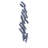
 Movie
Movie Controller


+ Open data

- Basic information
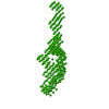
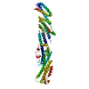
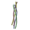
| Entry | Database: PDB / ID: 2b7m | ||||||
|---|---|---|---|---|---|---|---|
| Title | Crystal Structure of the S. cerevisiae Exocyst Component Exo70p | ||||||
 Components Components | Exocyst complex component EXO70 | ||||||
 Keywords Keywords | ENDOCYTOSIS/EXOCYTOSIS / exocyst / exocytosis / ENDOCYTOSIS-EXOCYTOSIS COMPLEX | ||||||
| Function / homology |  Function and homology information Function and homology informationexocyst assembly / exocyst localization / exocyst / prospore membrane / incipient cellular bud site / cellular bud tip / Golgi to plasma membrane transport / cellular bud neck / : / mating projection tip ...exocyst assembly / exocyst localization / exocyst / prospore membrane / incipient cellular bud site / cellular bud tip / Golgi to plasma membrane transport / cellular bud neck / : / mating projection tip / exocytosis / Rho protein signal transduction / transport vesicle / phosphatidylinositol-4,5-bisphosphate binding / small GTPase binding / protein transport / plasma membrane / cytoplasm Similarity search - Function | ||||||
| Biological species |  | ||||||
| Method |  X-RAY DIFFRACTION / SYNCHROTRON / MAD / Resolution: 3.5 Å X-RAY DIFFRACTION / SYNCHROTRON / MAD / Resolution: 3.5 Å | ||||||
 Authors Authors | Hamburger, Z.A. / Hamburger, A.E. / West, A.P. / Weis, W.I. | ||||||
 Citation Citation | Journal: J.Mol.Biol. / Year: 2006 Title: Crystal Structure of the S.cerevisiae Exocyst Component Exo70p Authors: Hamburger, Z.A. / Hamburger, A.E. / West, A.P. / Weis, W.I. | ||||||
| History |
|
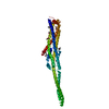
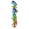
- Structure visualization
| Structure viewer | Molecule: MolmilJmol/JSmol |
|---|
- Downloads & links
-Download
| PDBx/mmCIF format | 2b7m.cif.gz | 380.7 KB | Display | PDBx/mmCIF format |
|---|---|---|---|---|
| PDB format | pdb2b7m.ent.gz | 317.9 KB | Display | PDB format |
| PDBx/mmJSON format | 2b7m.json.gz | Tree view | PDBx/mmJSON format | |
| Others |  Other downloads Other downloads |
-Validation report
| Arichive directory | https://data.pdbj.org/pub/pdb/validation_reports/b7/2b7mftp://data.pdbj.org/pub/pdb/validation_reports/b7/2b7m | HTTPS FTP |
|---|
-Related structure data
| Similar structure data |
|---|


-Links
 PDBj
PDBj
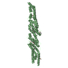
- Assembly
| Deposited unit | 
| ||||||||
|---|---|---|---|---|---|---|---|---|---|
| 1 | 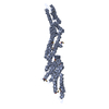
| ||||||||
| 2 | 
| ||||||||
| 3 | 
| ||||||||
| 4 | 
| ||||||||
| Unit cell |
|
-Components
| #1: Protein | Mass: 65537.406 Da / Num. of mol.: 4 Source method: isolated from a genetically manipulated source Source: (gene. exp.) Gene: EXO70 / Plasmid: pProEx-HTb / Production host:  Has protein modification | Y | |
|---|
-Experimental details
-Experiment
| Experiment | Method: X-RAY DIFFRACTION / Number of used crystals: 1 |
|---|
- Sample preparation
| Crystal | Density Matthews: 2.84 Å3/Da / Density % sol: 56.65 % |
|---|---|
| Crystal grow | Temperature: 295 K / Method: vapor diffusion, hanging drop / pH: 6.1 Details: 0.1 M MES pH 6.1, 20 % PEG 20000, 0.2 M magnesium acetate, VAPOR DIFFUSION, HANGING DROP, temperature 295K |
-Data collection
| Diffraction | Mean temperature: 100 K | ||||||||||||
|---|---|---|---|---|---|---|---|---|---|---|---|---|---|
| Diffraction source | Source: SYNCHROTRON / Site: ALS  / Beamline: 5.0.2 / Wavelength: 0.97950, 0.97963, 0.9393 / Beamline: 5.0.2 / Wavelength: 0.97950, 0.97963, 0.9393 | ||||||||||||
| Detector | Type: ADSC QUANTUM 210 / Detector: CCD / Date: Dec 5, 2004 | ||||||||||||
| Radiation | Monochromator: Si 111 CHANNEL / Protocol: MAD / Monochromatic (M) / Laue (L): M / Scattering type: x-ray | ||||||||||||
| Radiation wavelength |
| ||||||||||||
| Reflection | Resolution: 3.5→20 Å / Num. all: 36080 / Num. obs: 36080 / % possible obs: 95 % / Observed criterion σ(F): 2 / Observed criterion σ(I): 2 / Redundancy: 2.7 % / Rsym value: 0.076 / Net I/σ(I): 12.6 | ||||||||||||
| Reflection shell | Resolution: 3.5→3.62 Å / Redundancy: 3 % / Rsym value: 0.354 / % possible all: 92 |
- Processing
| Software |
| ||||||||||||||||||||||||||||||||||||||||||||||||||||||||||||
|---|---|---|---|---|---|---|---|---|---|---|---|---|---|---|---|---|---|---|---|---|---|---|---|---|---|---|---|---|---|---|---|---|---|---|---|---|---|---|---|---|---|---|---|---|---|---|---|---|---|---|---|---|---|---|---|---|---|---|---|---|---|
| Refinement | Method to determine structure: MAD / Resolution: 3.5→20 Å / Rfactor Rfree error: 0.007 / Data cutoff high absF: 4031626.48 / Data cutoff low absF: 0 / Isotropic thermal model: GROUP / Cross valid method: THROUGHOUT / σ(F): 0 / Stereochemistry target values: Engh & Huber
| ||||||||||||||||||||||||||||||||||||||||||||||||||||||||||||
| Solvent computation | Solvent model: FLAT MODEL / Bsol: 17.2292 Å2 / ksol: 0.157954 e/Å3 | ||||||||||||||||||||||||||||||||||||||||||||||||||||||||||||
| Displacement parameters | Biso mean: 129.2 Å2
| ||||||||||||||||||||||||||||||||||||||||||||||||||||||||||||
| Refine analyze |
| ||||||||||||||||||||||||||||||||||||||||||||||||||||||||||||
| Refinement step | Cycle: LAST / Resolution: 3.5→20 Å
| ||||||||||||||||||||||||||||||||||||||||||||||||||||||||||||
| Refine LS restraints |
| ||||||||||||||||||||||||||||||||||||||||||||||||||||||||||||
| Refine LS restraints NCS | NCS model details: CONSTR | ||||||||||||||||||||||||||||||||||||||||||||||||||||||||||||
| LS refinement shell | Resolution: 3.5→3.62 Å / Rfactor Rfree error: 0.029 / Total num. of bins used: 10
| ||||||||||||||||||||||||||||||||||||||||||||||||||||||||||||
| Xplor file | Serial no: 1 / Param file: protein_rep.param / Topol file: protein.top |
