 Movie
Movie Controller

[English] 日本語
 Yorodumi
- PDB-2b3l: Crystal structure of type I human methionine aminopeptidase in th... -
+ Open data

- Basic information
| Entry | Database: PDB / ID: 2b3l | ||||||
|---|---|---|---|---|---|---|---|
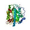
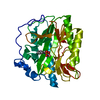
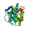
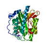
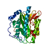
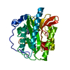
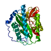
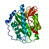
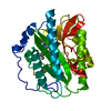
| Title | Crystal structure of type I human methionine aminopeptidase in the apo form | ||||||
 Components Components | Methionine aminopeptidase 1 | ||||||
 Keywords Keywords | HYDROLASE / methionine aminopeptidase / human / metalloprotease / pitabread fold | ||||||
| Function / homology |  Function and homology information Function and homology informationmethionyl aminopeptidase / initiator methionyl aminopeptidase activity / metalloexopeptidase activity / metalloaminopeptidase activity / aminopeptidase activity / protein maturation / platelet aggregation / regulation of translation / Inactivation, recovery and regulation of the phototransduction cascade / proteolysis ...methionyl aminopeptidase / initiator methionyl aminopeptidase activity / metalloexopeptidase activity / metalloaminopeptidase activity / aminopeptidase activity / protein maturation / platelet aggregation / regulation of translation / Inactivation, recovery and regulation of the phototransduction cascade / proteolysis / zinc ion binding / cytoplasm / cytosol Similarity search - Function | ||||||
| Biological species |  Homo sapiens (human) Homo sapiens (human) | ||||||
| Method |  X-RAY DIFFRACTION / SYNCHROTRON / MOLECULAR REPLACEMENT / Resolution: 1.5 Å X-RAY DIFFRACTION / SYNCHROTRON / MOLECULAR REPLACEMENT / Resolution: 1.5 Å | ||||||
 Authors Authors | Addlagatta, A. / Hu, X. / Liu, J.O. / Matthews, B.W. | ||||||
 Citation Citation | Journal: Biochemistry / Year: 2005 Title: Structural Basis for the Functional Differences between Type I and Type II Human Methionine Aminopeptidases(,). Authors: Addlagatta, A. / Hu, X. / Liu, J.O. / Matthews, B.W. | ||||||
| History |
|
- Structure visualization
| Structure viewer | Molecule: MolmilJmol/JSmol |
|---|
- Downloads & links
-Download
| PDBx/mmCIF format | 2b3l.cif.gz | 161.4 KB | Display | PDBx/mmCIF format |
|---|---|---|---|---|
| PDB format | pdb2b3l.ent.gz | 125.7 KB | Display | PDB format |
| PDBx/mmJSON format | 2b3l.json.gz | Tree view | PDBx/mmJSON format | |
| Others |  Other downloads Other downloads |
-Validation report
| Arichive directory | https://data.pdbj.org/pub/pdb/validation_reports/b3/2b3lftp://data.pdbj.org/pub/pdb/validation_reports/b3/2b3l | HTTPS FTP |
|---|
-Related structure data
| Related structure data |  2b3hC  2b3kC  1yj3S C: citing same article ( S: Starting model for refinement |
|---|---|
| Similar structure data |

-Links
 PDBj
PDBj


- Assembly
| Deposited unit | 
| ||||||||
|---|---|---|---|---|---|---|---|---|---|
| 1 |
| ||||||||
| Unit cell |
|
-Components
| #1: Protein | Mass: 36935.926 Da / Num. of mol.: 1 / Fragment: residues 81-384 Source method: isolated from a genetically manipulated source Source: (gene. exp.) Homo sapiens (human) / Gene: METAP1, KIAA0094 / Plasmid: pET15b / Species (production host): Escherichia coli / Production host:  | ||||
|---|---|---|---|---|---|
| #2: Chemical | ChemComp-K /   Mass: 39.098 Da / Num. of mol.: 1 / Source method: obtained synthetically / Formula: K Mass: 39.098 Da / Num. of mol.: 1 / Source method: obtained synthetically / Formula: K | ||||
| #3: Chemical |   Mass: 92.094 Da / Num. of mol.: 2 / Source method: obtained synthetically / Formula: C3H8O3 Mass: 92.094 Da / Num. of mol.: 2 / Source method: obtained synthetically / Formula: C3H8O3#4: Chemical | ChemComp-ACY / |   Mass: 60.052 Da / Num. of mol.: 1 / Source method: obtained synthetically / Formula: C2H4O2 Mass: 60.052 Da / Num. of mol.: 1 / Source method: obtained synthetically / Formula: C2H4O2#5: Water | ChemComp-HOH / |  Mass: 18.015 Da / Num. of mol.: 486 / Source method: isolated from a natural source / Formula: H2O Mass: 18.015 Da / Num. of mol.: 486 / Source method: isolated from a natural source / Formula: H2O |
-Experimental details
-Experiment
| Experiment | Method: X-RAY DIFFRACTION / Number of used crystals: 1 |
|---|
- Sample preparation
| Crystal | Density Matthews: 2.38 Å3/Da / Density % sol: 48.23 % |
|---|---|
| Crystal grow | Temperature: 298 K / Method: vapor diffusion, hanging drop / pH: 6 Details: PEG 2000, potassium chloride, hepes, sodium chloride, pH 6.0, VAPOR DIFFUSION, HANGING DROP, temperature 298K |
-Data collection
| Diffraction | Mean temperature: 100 K |
|---|---|
| Diffraction source | Source: SYNCHROTRON / Site: ALS  / Beamline: 8.2.1 / Wavelength: 1 Å / Beamline: 8.2.1 / Wavelength: 1 Å |
| Detector | Type: ADSC QUANTUM 210 / Detector: CCD / Date: Jul 6, 2004 / Details: Double Crystal Si(111) |
| Radiation | Monochromator: Double Crystal Si(111) / Protocol: SINGLE WAVELENGTH / Monochromatic (M) / Laue (L): M / Scattering type: x-ray |
| Radiation wavelength | Wavelength: 1 Å / Relative weight: 1 |
| Reflection | Resolution: 1.5→50 Å / Num. all: 51841 / Num. obs: 51841 / % possible obs: 95.1 % / Observed criterion σ(F): 0 / Observed criterion σ(I): 0 / Rmerge(I) obs: 0.031 / Χ2: 0.992 |
| Reflection shell | Resolution: 1.5→1.55 Å / % possible obs: 82.3 % / Rmerge(I) obs: 0.128 / Num. measured obs: 4470 / Χ2: 0.976 / % possible all: 83.3 |
- Processing
| Software |
| ||||||||||||||||||||||||||||
|---|---|---|---|---|---|---|---|---|---|---|---|---|---|---|---|---|---|---|---|---|---|---|---|---|---|---|---|---|---|
| Refinement | Method to determine structure: MOLECULAR REPLACEMENT Starting model: PDB Entry 1YJ3 Resolution: 1.5→50 Å / σ(F): 0 / σ(I): 0 / Stereochemistry target values: Engh & Huber
| ||||||||||||||||||||||||||||
| Displacement parameters | Biso mean: 25.8 Å2 | ||||||||||||||||||||||||||||
| Refinement step | Cycle: LAST / Resolution: 1.5→50 Å
|
