- PDB-2b31: Crystal structure of the complex formed between goat signalling p... -
+
Open data
ID or keywords:
Loading...
-
Basic information
Entry
Database: PDB / ID: 2b31
Title
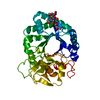
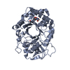
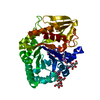
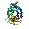
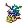
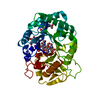
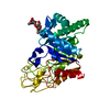
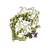
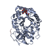
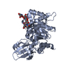
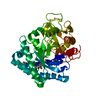
Crystal structure of the complex formed between goat signalling protein with pentasaccharide at 3.1 A resolution reveals large scale conformational changes in the residues of TIM barrel
Components
Chitinase-3-like protein 1, SPG-40
Keywords
SIGNALING PROTEIN / SIGNALLING PROTEIN / COMPLEX PENTASACCHARIDE
Function / homology
Function and homology information
response to interleukin-6 / activation of NF-kappaB-inducing kinase activity / positive regulation of peptidyl-threonine phosphorylation / chitin catabolic process / chitin binding / response to tumor necrosis factor / response to mechanical stimulus / lung development / response to interleukin-1 / positive regulation of interleukin-8 production ...response to interleukin-6 / activation of NF-kappaB-inducing kinase activity / positive regulation of peptidyl-threonine phosphorylation / chitin catabolic process / chitin binding / response to tumor necrosis factor / response to mechanical stimulus / lung development / response to interleukin-1 / positive regulation of interleukin-8 production / cellular response to tumor necrosis factor / positive regulation of angiogenesis / carbohydrate binding / carbohydrate metabolic process / positive regulation of ERK1 and ERK2 cascade / positive regulation of phosphatidylinositol 3-kinase/protein kinase B signal transduction / inflammatory response / apoptotic process / perinuclear region of cytoplasm / endoplasmic reticulum / : / cytoplasm Similarity search - Function
SEQUENCE The authors state that the aminoacid sequence in the database is incorrect for residues ...SEQUENCE The authors state that the aminoacid sequence in the database is incorrect for residues 54, 152, 226, 227 and 382. The authors state that residue 232 should be deleted from the sequence database reference.
Resolution: 3.1→56.7 Å / Cor.coef. Fo:Fc: 0.932 / Cor.coef. Fo:Fc free: 0.886 / SU B: 25.315 / SU ML: 0.464 / Cross valid method: THROUGHOUT / σ(F): 0 / ESU R Free: 0.424 / Stereochemistry target values: MAXIMUM LIKELIHOOD / Details: HYDROGENS HAVE BEEN ADDED IN THE RIDING POSITIONS
Rfactor
Num. reflection
% reflection
Selection details
Rfree
0.21782
403
4.7 %
RANDOM
Rwork
0.1766
-
-
-
obs
0.17859
8090
98.17 %
-
all
-
8493
-
-
Solvent computation
Ion probe radii: 0.8 Å / Shrinkage radii: 0.8 Å / VDW probe radii: 1.4 Å / Solvent model: BABINET MODEL WITH MASK
Displacement parameters
Biso mean: 24.973 Å2
Baniso -1
Baniso -2
Baniso -3
1-
0.4 Å2
0 Å2
0 Å2
2-
-
0.08 Å2
0 Å2
3-
-
-
-0.48 Å2
Refinement step
Cycle: LAST / Resolution: 3.1→56.7 Å
Protein
Nucleic acid
Ligand
Solvent
Total
Num. atoms
2876
0
98
71
3045
Refine LS restraints
Refine-ID
Type
Dev ideal
Dev ideal target
Number
X-RAY DIFFRACTION
r_bond_refined_d
0.02
0.021
3057
X-RAY DIFFRACTION
r_bond_other_d
0.003
0.02
2703
X-RAY DIFFRACTION
r_angle_refined_deg
2.44
1.972
4152
X-RAY DIFFRACTION
r_angle_other_deg
2.381
3
6234
X-RAY DIFFRACTION
r_dihedral_angle_1_deg
2.243
3
359
X-RAY DIFFRACTION
r_dihedral_angle_2_deg
X-RAY DIFFRACTION
r_dihedral_angle_3_deg
22.793
15
497
X-RAY DIFFRACTION
r_dihedral_angle_4_deg
X-RAY DIFFRACTION
r_chiral_restr
0.152
0.2
459
X-RAY DIFFRACTION
r_gen_planes_refined
0.007
0.02
3334
X-RAY DIFFRACTION
r_gen_planes_other
0.006
0.02
671
X-RAY DIFFRACTION
r_nbd_refined
0.296
0.3
744
X-RAY DIFFRACTION
r_nbd_other
0.268
0.3
2855
X-RAY DIFFRACTION
r_nbtor_refined
X-RAY DIFFRACTION
r_nbtor_other
0.472
0.5
10
X-RAY DIFFRACTION
r_xyhbond_nbd_refined
0.233
0.5
165
X-RAY DIFFRACTION
r_xyhbond_nbd_other
0.231
0.5
5
X-RAY DIFFRACTION
r_metal_ion_refined
X-RAY DIFFRACTION
r_metal_ion_other
X-RAY DIFFRACTION
r_symmetry_vdw_refined
0.311
0.3
6
X-RAY DIFFRACTION
r_symmetry_vdw_other
0.409
0.3
19
X-RAY DIFFRACTION
r_symmetry_hbond_refined
0.258
0.5
4
X-RAY DIFFRACTION
r_symmetry_hbond_other
X-RAY DIFFRACTION
r_symmetry_metal_ion_refined
X-RAY DIFFRACTION
r_symmetry_metal_ion_other
X-RAY DIFFRACTION
r_mcbond_it
1.683
1.5
1791
X-RAY DIFFRACTION
r_mcbond_other
X-RAY DIFFRACTION
r_mcangle_it
3.163
2
2872
X-RAY DIFFRACTION
r_scbond_it
3.417
3
1266
X-RAY DIFFRACTION
r_scangle_it
5.634
4.5
1280
X-RAY DIFFRACTION
r_rigid_bond_restr
X-RAY DIFFRACTION
r_sphericity_free
X-RAY DIFFRACTION
r_sphericity_bonded
LS refinement shell
Resolution: 3.1→3.18 Å / Total num. of bins used: 20 /
Rfactor
Num. reflection
Rfree
0.231
30
Rwork
0.243
609
+
About Yorodumi
-
News
-
Feb 9, 2022. New format data for meta-information of EMDB entries
New format data for meta-information of EMDB entries
Version 3 of the EMDB header file is now the official format.
The previous official version 1.9 will be removed from the archive.
In the structure databanks used in Yorodumi, some data are registered as the other names, "COVID-19 virus" and "2019-nCoV". Here are the details of the virus and the list of structure data.
Jan 31, 2019. EMDB accession codes are about to change! (news from PDBe EMDB page)
EMDB accession codes are about to change! (news from PDBe EMDB page)
The allocation of 4 digits for EMDB accession codes will soon come to an end. Whilst these codes will remain in use, new EMDB accession codes will include an additional digit and will expand incrementally as the available range of codes is exhausted. The current 4-digit format prefixed with “EMD-” (i.e. EMD-XXXX) will advance to a 5-digit format (i.e. EMD-XXXXX), and so on. It is currently estimated that the 4-digit codes will be depleted around Spring 2019, at which point the 5-digit format will come into force.
The EM Navigator/Yorodumi systems omit the EMD- prefix.
Related info.:Q: What is EMD? / ID/Accession-code notation in Yorodumi/EM Navigator
Yorodumi is a browser for structure data from EMDB, PDB, SASBDB, etc.
This page is also the successor to EM Navigator detail page, and also detail information page/front-end page for Omokage search.
The word "yorodu" (or yorozu) is an old Japanese word meaning "ten thousand". "mi" (miru) is to see.
Related info.:EMDB / PDB / SASBDB / Comparison of 3 databanks / Yorodumi Search / Aug 31, 2016. New EM Navigator & Yorodumi / Yorodumi Papers / Jmol/JSmol / Function and homology information / Changes in new EM Navigator and Yorodumi
 Movie
Movie Controller

 Yorodumi
 Open data
 Basic information
 Components
 Keywords
 Function and homology information
 X-RAY DIFFRACTION /
  Authors
 Citation
 Structure visualization
 Downloads & links
 Other downloads


 PDBj
PDBj Assembly
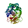
 Mass: 18.015 Da / Num. of mol.: 71 / Source method: isolated from a natural source / Formula: H2O
 Sample preparation
 Processing