 Movie
Movie Controller

[English] 日本語
 Yorodumi
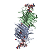
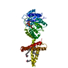
- PDB-2axo: X-Ray Crystal Structure of Protein AGR_C_4864 from Agrobacterium ... -
+ Open data

- Basic information
| Entry | Database: PDB / ID: 2axo | ||||||
|---|---|---|---|---|---|---|---|
| Title | X-Ray Crystal Structure of Protein AGR_C_4864 from Agrobacterium tumefaciens. Northeast Structural Genomics Consortium Target AtR35. | ||||||
 Components Components | hypothetical protein Atu2684 | ||||||
 Keywords Keywords | UNKNOWN FUNCTION / alpha beta protein. / Structural Genomics / PSI / Protein Structure Initiative / Northeast Structural Genomics Consortium / NESG | ||||||
| Function / homology | Protein of unknown function DUF1223 / Protein of unknown function (DUF1223) / Thioredoxin-like superfamily / DUF1223 domain-containing protein / :  Function and homology information Function and homology information | ||||||
| Biological species |  Agrobacterium tumefaciens str. (bacteria) Agrobacterium tumefaciens str. (bacteria) | ||||||
| Method |  X-RAY DIFFRACTION / SYNCHROTRON / SAD / Resolution: 1.8 Å X-RAY DIFFRACTION / SYNCHROTRON / SAD / Resolution: 1.8 Å | ||||||
 Authors Authors | Forouhar, F. / Abashidze, M. / Benach, J. / Xiao, R. / Janjua, H. / Conover, K. / Acton, T.B. / Montelione, G.T. / Tong, L. / Hunt, J.F. / Northeast Structural Genomics Consortium (NESG) | ||||||
 Citation Citation | Journal: To be Published Title: Crystal Structure of the Hypothetical Protein AGR_C_4864 from Agrobacterium tumefaciens, NESG target AtR35 Authors: Forouhar, F. / Abashidze, M. / Benach, J. / Xiao, R. / Janjua, H. / Conover, K. / Acton, T.B. / Montelione, G.T. / Tong, L. / Hunt, J.F. | ||||||
| History |
|
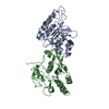
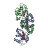
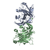
- Structure visualization
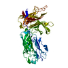
| Structure viewer | Molecule: MolmilJmol/JSmol |
|---|
- Downloads & links
-Download
| PDBx/mmCIF format | 2axo.cif.gz | 58.3 KB | Display | PDBx/mmCIF format |
|---|---|---|---|---|
| PDB format | pdb2axo.ent.gz | 41.9 KB | Display | PDB format |
| PDBx/mmJSON format | 2axo.json.gz | Tree view | PDBx/mmJSON format | |
| Others |  Other downloads Other downloads |
-Validation report
| Arichive directory | https://data.pdbj.org/pub/pdb/validation_reports/ax/2axoftp://data.pdbj.org/pub/pdb/validation_reports/ax/2axo | HTTPS FTP |
|---|
-Related structure data
| Similar structure data | |
|---|---|
| Other databases |
-Links
 PDBj
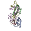
PDBj- Assembly
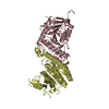
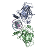
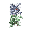
| Deposited unit | 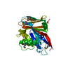
| ||||||||
|---|---|---|---|---|---|---|---|---|---|
| 1 | 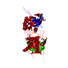
| ||||||||
| Unit cell |
| ||||||||
| Details | possibly dimer. |
-Components
| #1: Protein | Mass: 29822.830 Da / Num. of mol.: 1 Source method: isolated from a genetically manipulated source Source: (gene. exp.) Agrobacterium tumefaciens str. (bacteria)Species: Agrobacterium tumefaciens / Strain: C58 / Gene: MDK4.6 / Plasmid: BL21 / Production host: |
|---|---|
| #2: Water | ChemComp-HOH /  Mass: 18.015 Da / Num. of mol.: 116 / Source method: isolated from a natural source / Formula: H2O Mass: 18.015 Da / Num. of mol.: 116 / Source method: isolated from a natural source / Formula: H2O |
| Has protein modification | Y |
-Experimental details
-Experiment
| Experiment | Method: X-RAY DIFFRACTION / Number of used crystals: 1 |
|---|
- Sample preparation
| Crystal | Density Matthews: 1.6 Å3/Da / Density % sol: 23.1 % |
|---|---|
| Crystal grow | Temperature: 293 K / Method: vapor diffusion, hanging drop / pH: 7.5 Details: 20 mM Tris (pH 7.5), 18% PEG 2kMME, 200 mM sodium formate, and 5 mM DTT., VAPOR DIFFUSION, HANGING DROP, temperature 293K |
-Data collection
| Diffraction | Mean temperature: 100 K |
|---|---|
| Diffraction source | Source: SYNCHROTRON / Site: NSLS  / Beamline: X4A / Wavelength: 0.97923 Å / Beamline: X4A / Wavelength: 0.97923 Å |
| Detector | Type: ADSC QUANTUM 4 / Detector: CCD / Date: Aug 24, 2005 / Details: mirrors |
| Radiation | Monochromator: Si 111 CHANNEL / Protocol: SINGLE WAVELENGTH / Monochromatic (M) / Laue (L): M / Scattering type: x-ray |
| Radiation wavelength | Wavelength: 0.97923 Å / Relative weight: 1 |
| Reflection | Resolution: 1.72→20 Å / Num. all: 37482 / Num. obs: 37295 / % possible obs: 99.9 % / Observed criterion σ(F): 0 / Observed criterion σ(I): 0 / Redundancy: 6.6 % / Biso Wilson estimate: 10.5 Å2 / Rmerge(I) obs: 0.084 / Rsym value: 0.064 / Net I/σ(I): 23.4 |
| Reflection shell | Resolution: 1.72→1.75 Å / Redundancy: 4.6 % / Rmerge(I) obs: 0.452 / Mean I/σ(I) obs: 2.3 / Num. unique all: 1837 / Rsym value: 0.429 / % possible all: 99.8 |
- Processing
| Software |
| |||||||||||||||||||||||||
|---|---|---|---|---|---|---|---|---|---|---|---|---|---|---|---|---|---|---|---|---|---|---|---|---|---|---|
| Refinement | Method to determine structure: SAD / Resolution: 1.8→20 Å / Rfactor Rfree error: 0.005 / Data cutoff high absF: 228678.5 / Data cutoff low absF: 0 / Isotropic thermal model: OVERALL / Cross valid method: THROUGHOUT / σ(F): 2 / σ(I): 2 / Stereochemistry target values: Engh & Huber Details: XtalView and SnB were also used in the refinement and solution of this structure.
| |||||||||||||||||||||||||
| Solvent computation | Solvent model: FLAT MODEL / Bsol: 43.9 Å2 / ksol: 0.36423 e/Å3 | |||||||||||||||||||||||||
| Displacement parameters | Biso mean: 19.5 Å2
| |||||||||||||||||||||||||
| Refine analyze |
| |||||||||||||||||||||||||
| Refinement step | Cycle: LAST / Resolution: 1.8→20 Å
| |||||||||||||||||||||||||
| Refine LS restraints |
| |||||||||||||||||||||||||
| LS refinement shell | Resolution: 1.8→1.91 Å / Rfactor Rfree error: 0.013 / Total num. of bins used: 6
|
