 Movie
Movie Controller

[English] 日本語
 Yorodumi
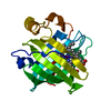
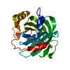
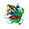
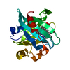
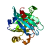
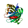
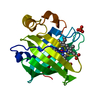
- PDB-2at0: 1.00 A Crystal Structure Of L133V Mutant of Nitrophorin 4 From Rh... -
+ Open data

- Basic information
| Entry | Database: PDB / ID: 2at0 | ||||||
|---|---|---|---|---|---|---|---|
| Title | 1.00 A Crystal Structure Of L133V Mutant of Nitrophorin 4 From Rhodnius Prolixus Complexed With Nitric Oxide at pH 5.6 | ||||||
 Components Components | Nitrophorin 4 | ||||||
 Keywords Keywords | TRANSPORT PROTEIN / Lipocalin / beta barrel / ferrous heme / nitric oxide | ||||||
| Function / homology |  Function and homology information Function and homology informationnitrite dismutase / histamine binding / nitric oxide binding / vasodilation / oxidoreductase activity / extracellular region / metal ion binding Similarity search - Function | ||||||
| Biological species |   Rhodnius prolixus (insect) Rhodnius prolixus (insect) | ||||||
| Method |  X-RAY DIFFRACTION / SYNCHROTRON / FOURIER SYNTHESIS / Resolution: 1 Å X-RAY DIFFRACTION / SYNCHROTRON / FOURIER SYNTHESIS / Resolution: 1 Å | ||||||
 Authors Authors | Amoia, A.M. / Montfort, W.R. | ||||||
 Citation Citation | Journal: To be Published Title: Heme distortion in nitrophorin 4: high resolution structures of mutated positions L123V and L133V and heme altered proteins Authors: Amoia, A.M. / Montfort, W.R. | ||||||
| History |
| ||||||
| Remark 600 | HETEROGEN The heme is disordered by a rotation of 180 degrees around the CHA-Fe-CHC axis, which is ...HETEROGEN The heme is disordered by a rotation of 180 degrees around the CHA-Fe-CHC axis, which is reflected in conformation A and conformation D of the ligand. |
- Structure visualization
| Structure viewer | Molecule: MolmilJmol/JSmol |
|---|
- Downloads & links
-Download
| PDBx/mmCIF format | 2at0.cif.gz | 112.5 KB | Display | PDBx/mmCIF format |
|---|---|---|---|---|
| PDB format | pdb2at0.ent.gz | 86.5 KB | Display | PDB format |
| PDBx/mmJSON format | 2at0.json.gz | Tree view | PDBx/mmJSON format | |
| Others |  Other downloads Other downloads |
-Validation report
| Arichive directory | https://data.pdbj.org/pub/pdb/validation_reports/at/2at0ftp://data.pdbj.org/pub/pdb/validation_reports/at/2at0 | HTTPS FTP |
|---|
-Related structure data
| Related structure data |  2at3C 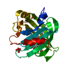 2at5C  2at6C 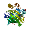 2at8C 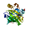 1x8oS C: citing same article ( S: Starting model for refinement |
|---|---|
| Similar structure data |

-Links
 PDBj
PDBj


- Assembly
| Deposited unit | 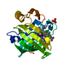
| |||||||||||||||
|---|---|---|---|---|---|---|---|---|---|---|---|---|---|---|---|---|
| 1 |
| |||||||||||||||
| Unit cell |
| |||||||||||||||
| Components on special symmetry positions |
| |||||||||||||||
| Details | The biological assembly is a monomer consisting of chain X and the heme, and can be generated by the identity operation: x,y,z |
-Components
| #1: Protein | Mass: 20278.637 Da / Num. of mol.: 1 / Mutation: L133V Source method: isolated from a genetically manipulated source Source: (gene. exp.) Rhodnius prolixus (insect) / Plasmid: pET17b / Species (production host): Escherichia coli / Production host:  |
|---|---|
| #2: Chemical | ChemComp-PO4 /   Mass: 94.971 Da / Num. of mol.: 1 / Source method: obtained synthetically / Formula: PO4 Mass: 94.971 Da / Num. of mol.: 1 / Source method: obtained synthetically / Formula: PO4 |
| #3: Chemical | ChemComp-HEM /   Mass: 616.487 Da / Num. of mol.: 1 / Source method: obtained synthetically / Formula: C34H32FeN4O4 Mass: 616.487 Da / Num. of mol.: 1 / Source method: obtained synthetically / Formula: C34H32FeN4O4 |
| #4: Chemical | ChemComp-NO /   Mass: 30.006 Da / Num. of mol.: 1 / Source method: obtained synthetically / Formula: NO Mass: 30.006 Da / Num. of mol.: 1 / Source method: obtained synthetically / Formula: NO |
| #5: Water | ChemComp-HOH /  Mass: 18.015 Da / Num. of mol.: 284 / Source method: isolated from a natural source / Formula: H2O Mass: 18.015 Da / Num. of mol.: 284 / Source method: isolated from a natural source / Formula: H2O |
| Has protein modification | Y |
-Experimental details
-Experiment
| Experiment | Method: X-RAY DIFFRACTION / Number of used crystals: 1 |
|---|
- Sample preparation
| Crystal | Density Matthews: 1.62 Å3/Da / Density % sol: 23.63 % |
|---|---|
| Crystal grow | Temperature: 300 K / Method: vapor diffusion, hanging drop / pH: 5.6 Details: ammonium phosphate, pH 5.6, VAPOR DIFFUSION, HANGING DROP, temperature 300.0K |
-Data collection
| Diffraction | Mean temperature: 100 K |
|---|---|
| Diffraction source | Source: SYNCHROTRON / Site: SSRL  / Beamline: BL9-2 / Wavelength: 0.98 Å / Beamline: BL9-2 / Wavelength: 0.98 Å |
| Detector | Type: MARMOSAIC 325 mm CCD / Detector: CCD / Date: Jul 20, 2005 |
| Radiation | Monochromator: double crystal / Protocol: SINGLE WAVELENGTH / Monochromatic (M) / Laue (L): M / Scattering type: x-ray |
| Radiation wavelength | Wavelength: 0.98 Å / Relative weight: 1 |
| Reflection | Resolution: 1→36.51 Å / Num. obs: 83901 / % possible obs: 99.8 % / Observed criterion σ(F): 0 / Observed criterion σ(I): 0 / Redundancy: 4.1 % / Biso Wilson estimate: 9.3 Å2 / Rmerge(I) obs: 0.076 / Rsym value: 0.076 / Net I/σ(I): 13.9 |
| Reflection shell | Resolution: 1→1.05 Å / % possible obs: 98.7 % / Redundancy: 3.4 % / Rmerge(I) obs: 0.178 / Mean I/σ(I) obs: 5.3 / Num. measured obs: 12069 / Num. unique all: 12069 / Rsym value: 0.178 / % possible all: 98 |
- Processing
| Software |
| ||||||||||||||||||||||||||||||||||||||||||||||||||||||||||||||||||||||||||||||||||||||||||||||||||||||||||||||
|---|---|---|---|---|---|---|---|---|---|---|---|---|---|---|---|---|---|---|---|---|---|---|---|---|---|---|---|---|---|---|---|---|---|---|---|---|---|---|---|---|---|---|---|---|---|---|---|---|---|---|---|---|---|---|---|---|---|---|---|---|---|---|---|---|---|---|---|---|---|---|---|---|---|---|---|---|---|---|---|---|---|---|---|---|---|---|---|---|---|---|---|---|---|---|---|---|---|---|---|---|---|---|---|---|---|---|---|---|---|---|---|
| Refinement | Method to determine structure: FOURIER SYNTHESIS Starting model: pdb entry 1X8O Resolution: 1→36.47 Å / Cor.coef. Fo:Fc: 0.97 / Cor.coef. Fo:Fc free: 0.963 / SU B: 0.514 / SU ML: 0.013 Isotropic thermal model: anisotropic; residues 32-35 isotropic due to lack of density Cross valid method: FREE R / σ(F): 0 / σ(I): 0 / ESU R: 0.026 / ESU R Free: 0.026 / Stereochemistry target values: Engh & Huber Details: Anisotropic refinment reduced Free R. HYDROGENS HAVE BEEN ADDED IN THE RIDING POSITIONS
| ||||||||||||||||||||||||||||||||||||||||||||||||||||||||||||||||||||||||||||||||||||||||||||||||||||||||||||||
| Solvent computation | Ion probe radii: 0.8 Å / Shrinkage radii: 0.8 Å / VDW probe radii: 1.2 Å / Solvent model: MASK | ||||||||||||||||||||||||||||||||||||||||||||||||||||||||||||||||||||||||||||||||||||||||||||||||||||||||||||||
| Displacement parameters | Biso mean: 12.753 Å2
| ||||||||||||||||||||||||||||||||||||||||||||||||||||||||||||||||||||||||||||||||||||||||||||||||||||||||||||||
| Refine analyze | Luzzati coordinate error obs: 0.28 Å / Luzzati d res low obs: 4 Å | ||||||||||||||||||||||||||||||||||||||||||||||||||||||||||||||||||||||||||||||||||||||||||||||||||||||||||||||
| Refinement step | Cycle: LAST / Resolution: 1→36.47 Å
| ||||||||||||||||||||||||||||||||||||||||||||||||||||||||||||||||||||||||||||||||||||||||||||||||||||||||||||||
| Refine LS restraints |
| ||||||||||||||||||||||||||||||||||||||||||||||||||||||||||||||||||||||||||||||||||||||||||||||||||||||||||||||
| LS refinement shell | Resolution: 1→1.026 Å / Total num. of bins used: 20
|
