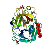
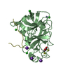
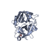
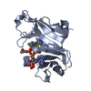
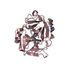
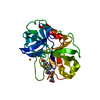
regulation of cAMP-dependent protein kinase activity / Signaling by MST1 / regulation of receptor signaling pathway via JAK-STAT / negative regulation of gluconeogenesis / receptor tyrosine kinase binding / : / proteolysis / extracellular space / extracellular region Similarity search - Function
Macrophage stimulating protein / Hepatocyte growth factor/Macrophage stimulatory protein / divergent subfamily of APPLE domains / : / PAN/Apple domain profile. / PAN domain / PAN/Apple domain / Kringle domain / Kringle / Kringle, conserved site ...Macrophage stimulating protein / Hepatocyte growth factor/Macrophage stimulatory protein / divergent subfamily of APPLE domains / : / PAN/Apple domain profile. / PAN domain / PAN/Apple domain / Kringle domain / Kringle / Kringle, conserved site / Kringle superfamily / Kringle domain signature. / Kringle domain profile. / Kringle domain / Kringle-like fold / Peptidase S1A, chymotrypsin family / Serine proteases, trypsin domain profile. / Trypsin-like serine protease / Serine proteases, trypsin domain / Trypsin / Trypsin-like serine proteases / Thrombin, subunit H / Peptidase S1, PA clan, chymotrypsin-like fold / Peptidase S1, PA clan / Beta Barrel / Mainly Beta Similarity search - Domain/homology
Resolution: 1.85→50 Å / Cor.coef. Fo:Fc: 0.948 / Cor.coef. Fo:Fc free: 0.929 / Cross valid method: THROUGHOUT / ESU R Free: 0.129 / Stereochemistry target values: MAXIMUM LIKELIHOOD / Details: HYDROGENS HAVE BEEN ADDED IN THE RIDING POSITIONS
Rfactor
Num. reflection
% reflection
Selection details
Rfree
0.23678
1221
5.1 %
RANDOM
Rwork
0.19802
-
-
-
all
0.1999
22661
-
-
obs
0.1999
22661
97.82 %
-
Solvent computation
Ion probe radii: 0.8 Å / Shrinkage radii: 0.8 Å / VDW probe radii: 1.4 Å / Solvent model: BABINET MODEL WITH MASK
Displacement parameters
Biso mean: 25.129 Å2
Baniso -1
Baniso -2
Baniso -3
1-
-0.9 Å2
0 Å2
0 Å2
2-
-
1.08 Å2
0 Å2
3-
-
-
-0.18 Å2
Refinement step
Cycle: LAST / Resolution: 1.85→50 Å
Protein
Nucleic acid
Ligand
Solvent
Total
Num. atoms
1730
0
0
155
1885
Refine LS restraints
Refine-ID
Type
Dev ideal
Dev ideal target
Number
X-RAY DIFFRACTION
r_bond_refined_d
0.017
0.021
1780
X-RAY DIFFRACTION
r_bond_other_d
0.002
0.02
1620
X-RAY DIFFRACTION
r_angle_refined_deg
1.599
1.943
2427
X-RAY DIFFRACTION
r_angle_other_deg
0.855
3
3760
X-RAY DIFFRACTION
r_dihedral_angle_1_deg
6.169
5
227
X-RAY DIFFRACTION
r_chiral_restr
0.099
0.2
270
X-RAY DIFFRACTION
r_gen_planes_refined
0.007
0.02
1987
X-RAY DIFFRACTION
r_gen_planes_other
0.003
0.02
355
X-RAY DIFFRACTION
r_nbd_refined
0.186
0.2
311
X-RAY DIFFRACTION
r_nbd_other
0.241
0.2
1897
X-RAY DIFFRACTION
r_nbtor_other
0.083
0.2
1035
X-RAY DIFFRACTION
r_xyhbond_nbd_refined
0.219
0.2
97
X-RAY DIFFRACTION
r_symmetry_vdw_refined
0.041
0.2
2
X-RAY DIFFRACTION
r_symmetry_vdw_other
0.29
0.2
28
X-RAY DIFFRACTION
r_symmetry_hbond_refined
0.156
0.2
11
X-RAY DIFFRACTION
r_mcbond_it
3.36
5
1135
X-RAY DIFFRACTION
r_mcangle_it
4.352
6
1827
X-RAY DIFFRACTION
r_scbond_it
3.853
5
645
X-RAY DIFFRACTION
r_scangle_it
5.502
7.5
600
LS refinement shell
Resolution: 1.845→1.893 Å / Total num. of bins used: 20
Rfactor
Num. reflection
Rfree
0.282
74
Rwork
0.236
1515
obs
-
1515
+
About Yorodumi
-
News
-
Feb 9, 2022. New format data for meta-information of EMDB entries
New format data for meta-information of EMDB entries
Version 3 of the EMDB header file is now the official format.
The previous official version 1.9 will be removed from the archive.
In the structure databanks used in Yorodumi, some data are registered as the other names, "COVID-19 virus" and "2019-nCoV". Here are the details of the virus and the list of structure data.
Jan 31, 2019. EMDB accession codes are about to change! (news from PDBe EMDB page)
EMDB accession codes are about to change! (news from PDBe EMDB page)
The allocation of 4 digits for EMDB accession codes will soon come to an end. Whilst these codes will remain in use, new EMDB accession codes will include an additional digit and will expand incrementally as the available range of codes is exhausted. The current 4-digit format prefixed with “EMD-” (i.e. EMD-XXXX) will advance to a 5-digit format (i.e. EMD-XXXXX), and so on. It is currently estimated that the 4-digit codes will be depleted around Spring 2019, at which point the 5-digit format will come into force.
The EM Navigator/Yorodumi systems omit the EMD- prefix.
Related info.:Q: What is EMD? / ID/Accession-code notation in Yorodumi/EM Navigator
Yorodumi is a browser for structure data from EMDB, PDB, SASBDB, etc.
This page is also the successor to EM Navigator detail page, and also detail information page/front-end page for Omokage search.
The word "yorodu" (or yorozu) is an old Japanese word meaning "ten thousand". "mi" (miru) is to see.
Related info.:EMDB / PDB / SASBDB / Comparison of 3 databanks / Yorodumi Search / Aug 31, 2016. New EM Navigator & Yorodumi / Yorodumi Papers / Jmol/JSmol / Function and homology information / Changes in new EM Navigator and Yorodumi
 Movie
Movie Controller


 Open data
 Basic information
 Components
 Keywords
 Function and homology information
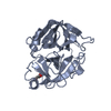
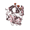
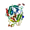
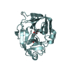
 Homo sapiens (human)
 X-RAY DIFFRACTION /
  Authors
 Citation
 Structure visualization
 Downloads & links
 Other downloads
 PDBj
PDBj

 Assembly
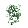

 Mass: 18.015 Da / Num. of mol.: 155 / Source method: isolated from a natural source / Formula: H2O
 Sample preparation
 / Beamline: ID13 / Wavelength: 0.979 Å
 Processing