Resolution: 1.9→51.7 Å / Cor.coef. Fo:Fc: 0.959 / Cor.coef. Fo:Fc free: 0.944 / SU B: 7.909 / SU ML: 0.117 / TLS residual ADP flag: LIKELY RESIDUAL / Cross valid method: THROUGHOUT / ESU R: 0.147 / ESU R Free: 0.137 Stereochemistry target values: MAXIMUM LIKELIHOOD WITH PHASES Details: 1. HYDROGENS HAVE BEEN ADDED IN THE RIDING POSITIONS 2. DENSITY IS WEAK FOR RESIDUES 99-100. NCS WAS USED TO MODEL THIS REGION. (3) RESIDUES 147-148 WERE OMITTED FROM ALL CHAINS DUE TO POOR ...Details: 1. HYDROGENS HAVE BEEN ADDED IN THE RIDING POSITIONS 2. DENSITY IS WEAK FOR RESIDUES 99-100. NCS WAS USED TO MODEL THIS REGION. (3) RESIDUES 147-148 WERE OMITTED FROM ALL CHAINS DUE TO POOR DENSITY. 4. THE COORDINATION OF ALL FOUR ZINC IONS ARE EQUIVALENT. HOWEVER, THE ENVIRONMENT AROUND THE ZINC ION IN CHAIN D DISTORTS DURING REFINEMENT. THE STRUCTURE WAS INITIALLY SOLVED BY MOLECULAR REPLACEMENT USING THE 1EFZ SEARCH MODEL AND REFINED AGAINST THE 1.9 A NATIVE DATA. DURING REFINEMENT, 2.1 A DATA FROM A MAD EXPERIMENT AT THE ZINC EDGE WERE USED TO CALCULATE EXPERIMENTAL PHASES. THE SUBSTRUCTURE COULD BE SOLVED DE NOVO USING SHELXD AND THE PHASES WERE REFINED WITH AUTOSHARP. THESE PHASES WERE EXTENDED WITH DM USING AMPLITUDES FROM HIGHER RESOLUTION (1.9 A) NATIVE DATA SET. THESE DENSITY MODIFIED ZN-MAD PHASES WERE USED AS RESTRAINTS IN SUBSEQUENT REFINEMENT CYCLES AGAINST THE 1.9 A NATIVE DATA AND WERE INCLUDED IN THE FINAL REFINEMENT.
Rfactor
Num. reflection
% reflection
Selection details
Rfree
0.229
6626
5 %
RANDOM
Rwork
0.193
-
-
-
all
0.195
-
-
-
obs
-
124830
96.9 %
-
Solvent computation
Ion probe radii: 0.8 Å / Shrinkage radii: 0.8 Å / VDW probe radii: 1.2 Å / Solvent model: MASK
In the structure databanks used in Yorodumi, some data are registered as the other names, "COVID-19 virus" and "2019-nCoV". Here are the details of the virus and the list of structure data.
Jan 31, 2019. EMDB accession codes are about to change! (news from PDBe EMDB page)
EMDB accession codes are about to change! (news from PDBe EMDB page)
The allocation of 4 digits for EMDB accession codes will soon come to an end. Whilst these codes will remain in use, new EMDB accession codes will include an additional digit and will expand incrementally as the available range of codes is exhausted. The current 4-digit format prefixed with “EMD-” (i.e. EMD-XXXX) will advance to a 5-digit format (i.e. EMD-XXXXX), and so on. It is currently estimated that the 4-digit codes will be depleted around Spring 2019, at which point the 5-digit format will come into force.
The EM Navigator/Yorodumi systems omit the EMD- prefix.
Related info.:Q: What is EMD? / ID/Accession-code notation in Yorodumi/EM Navigator
Yorodumi is a browser for structure data from EMDB, PDB, SASBDB, etc.
This page is also the successor to EM Navigator detail page, and also detail information page/front-end page for Omokage search.
The word "yorodu" (or yorozu) is an old Japanese word meaning "ten thousand". "mi" (miru) is to see.
Related info.:EMDB / PDB / SASBDB / Comparison of 3 databanks / Yorodumi Search / Aug 31, 2016. New EM Navigator & Yorodumi / Yorodumi Papers / Jmol/JSmol / Function and homology information / Changes in new EM Navigator and Yorodumi
 Movie
Movie Controller

 Yorodumi
 Open data
 Basic information
 Components
 Keywords
 Function and homology information
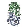
 Thermotoga maritima (bacteria)
 X-RAY DIFFRACTION /
  Authors
 Citation
 Structure visualization
 Downloads & links
 Other downloads


 PDBj
PDBj Assembly
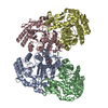


 Mass: 65.409 Da / Num. of mol.: 4 / Source method: obtained synthetically / Formula: Zn
 Mass: 35.453 Da / Num. of mol.: 5 / Source method: obtained synthetically / Formula: Cl
 Mass: 62.068 Da / Num. of mol.: 10 / Source method: obtained synthetically / Formula: C2H6O2
 Mass: 18.015 Da / Num. of mol.: 655 / Source method: isolated from a natural source / Formula: H2O
 Sample preparation
 Processing