 Movie
Movie Controller


+ Open data

- Basic information
| Entry | Database: PDB / ID: 2aps | ||||||
|---|---|---|---|---|---|---|---|
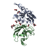
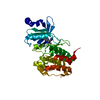
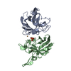
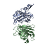
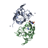
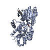
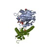
| Title | CU/ZN SUPEROXIDE DISMUTASE FROM ACTINOBACILLUS PLEUROPNEUMONIAE | ||||||
 Components Components | PROTEIN (CU,ZN SUPEROXIDE DISMUTASE) | ||||||
 Keywords Keywords | SUPEROXIDE DISMUTASE / SOD / WATER-MEDIATED DIMER / BETA BARREL | ||||||
| Function / homology |  Function and homology information Function and homology informationsuperoxide dismutase / superoxide dismutase activity / periplasmic space / copper ion binding Similarity search - Function | ||||||
| Biological species |  Actinobacillus pleuropneumoniae (bacteria) Actinobacillus pleuropneumoniae (bacteria) | ||||||
| Method |  X-RAY DIFFRACTION / SYNCHROTRON / MOLECULAR REPLACEMENT / Resolution: 1.9 Å X-RAY DIFFRACTION / SYNCHROTRON / MOLECULAR REPLACEMENT / Resolution: 1.9 Å | ||||||
 Authors Authors | Forest, K.T. / Langford, P.R. / Kroll, J.S. / Getzoff, E.D. | ||||||
 Citation Citation | Journal: J.Mol.Biol. / Year: 2000 Title: Cu,Zn superoxide dismutase structure from a microbial pathogen establishes a class with a conserved dimer interface. Authors: Forest, K.T. / Langford, P.R. / Kroll, J.S. / Getzoff, E.D. | ||||||
| History |
|
- Structure visualization
| Structure viewer | Molecule: MolmilJmol/JSmol |
|---|
- Downloads & links
-Download
| PDBx/mmCIF format | 2aps.cif.gz | 74 KB | Display | PDBx/mmCIF format |
|---|---|---|---|---|
| PDB format | pdb2aps.ent.gz | 54.9 KB | Display | PDB format |
| PDBx/mmJSON format | 2aps.json.gz | Tree view | PDBx/mmJSON format | |
| Others |  Other downloads Other downloads |
-Validation report
| Arichive directory | https://data.pdbj.org/pub/pdb/validation_reports/ap/2apsftp://data.pdbj.org/pub/pdb/validation_reports/ap/2aps | HTTPS FTP |
|---|
-Related structure data
| Related structure data | 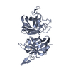 1yaiS S: Starting model for refinement |
|---|---|
| Similar structure data |

-Links
 PDBj
PDBj
- Assembly
| Deposited unit | 
| ||||||||
|---|---|---|---|---|---|---|---|---|---|
| 1 |
| ||||||||
| Unit cell |
| ||||||||
| Components on special symmetry positions |
| ||||||||
| Noncrystallographic symmetry (NCS) | NCS oper: (Code: given Matrix: (-0.859, 0.5063, -0.0765), Vector: |
-Components
| #1: Protein | Mass: 17254.371 Da / Num. of mol.: 2 Source method: isolated from a genetically manipulated source Details: EACH ACTIVE SITE CONTAINS ONE COPPER AND ONE ZINC. Source: (gene. exp.) Actinobacillus pleuropneumoniae (bacteria)Cellular location: PERIPLASM / Gene: SODC / Plasmid: PJSK157 / Cellular location (production host): PERIPLASM / Gene (production host): SODC / Production host: #2: Chemical | ChemComp-CU /   Mass: 63.546 Da / Num. of mol.: 4 / Source method: obtained synthetically / Formula: Cu Mass: 63.546 Da / Num. of mol.: 4 / Source method: obtained synthetically / Formula: Cu#3: Chemical |   Mass: 65.409 Da / Num. of mol.: 2 / Source method: obtained synthetically / Formula: Zn Mass: 65.409 Da / Num. of mol.: 2 / Source method: obtained synthetically / Formula: Zn#4: Water | ChemComp-HOH / |  Mass: 18.015 Da / Num. of mol.: 126 / Source method: isolated from a natural source / Formula: H2O Mass: 18.015 Da / Num. of mol.: 126 / Source method: isolated from a natural source / Formula: H2OHas protein modification | Y | Nonpolymer details | CU 300 AND CU 500 FORM CRYSTAL CONTACTS. CU 300 LIES ON A SPECIAL POSITION. CU 402 AND CU 602 ARE ...CU 300 AND CU 500 FORM CRYSTAL CONTACTS. CU 300 LIES ON A SPECIAL POSITION. CU 402 AND CU 602 ARE IN ACTIVE SITES. BOTH ZINC IONS ARE IN ACTIVE SITES. | Sequence details | THERE IS NO LEADER PEPTIDE IN THE PURIFIED PROTEIN (23 RESIDUES ARE CLEAVED) AND THE FIRST 5 ...THERE IS NO LEADER PEPTIDE IN THE PURIFIED PROTEIN (23 RESIDUES ARE CLEAVED) AND THE FIRST 5 RESIDUES OF THE EXPRESSED (MATURE) PROTEIN HAVE APPARENTLY | |
|---|
-Experimental details
-Experiment
| Experiment | Method: X-RAY DIFFRACTION / Number of used crystals: 1 |
|---|
- Sample preparation
| Crystal | Density Matthews: 2.8 Å3/Da / Density % sol: 42 % / Description: SEARCH MODEL WAS A DIMER. | ||||||||||||||||||||||||||||||
|---|---|---|---|---|---|---|---|---|---|---|---|---|---|---|---|---|---|---|---|---|---|---|---|---|---|---|---|---|---|---|---|
| Crystal grow | pH: 5 / Details: pH 5.0 | ||||||||||||||||||||||||||||||
| Crystal grow | *PLUS Method: vapor diffusion, hanging drop | ||||||||||||||||||||||||||||||
| Components of the solutions | *PLUS
|
-Data collection
| Diffraction | Mean temperature: 100 K |
|---|---|
| Diffraction source | Source: SYNCHROTRON / Site: SSRL  / Beamline: BL7-1 / Wavelength: 1.08 / Beamline: BL7-1 / Wavelength: 1.08 |
| Detector | Type: MARRESEARCH / Detector: IMAGE PLATE / Date: Jul 1, 1997 |
| Radiation | Protocol: SINGLE WAVELENGTH / Monochromatic (M) / Laue (L): M / Scattering type: x-ray |
| Radiation wavelength | Wavelength: 1.08 Å / Relative weight: 1 |
| Reflection | Resolution: 1.9→20 Å / Num. obs: 30234 / % possible obs: 98 % / Observed criterion σ(I): 0 / Redundancy: 3.5 % / Biso Wilson estimate: 27.6 Å2 / Rsym value: 0.054 / Net I/σ(I): 28 |
| Reflection shell | Resolution: 1.9→1.96 Å / Rmerge(I) obs: 0.092 / Mean I/σ(I) obs: 13.7 / % possible all: 92 |
| Reflection | *PLUS Num. measured all: 106143 / Rmerge(I) obs: 0.047 |
| Reflection shell | *PLUS % possible obs: 92 % |
- Processing
| Software |
| ||||||||||||||||||||||||||||||||||||||||||||||||||||||||||||||||||||||||||||||||
|---|---|---|---|---|---|---|---|---|---|---|---|---|---|---|---|---|---|---|---|---|---|---|---|---|---|---|---|---|---|---|---|---|---|---|---|---|---|---|---|---|---|---|---|---|---|---|---|---|---|---|---|---|---|---|---|---|---|---|---|---|---|---|---|---|---|---|---|---|---|---|---|---|---|---|---|---|---|---|---|---|---|
| Refinement | Method to determine structure: MOLECULAR REPLACEMENT Starting model: PDB ENTRY 1YAI Resolution: 1.9→20 Å / Data cutoff high absF: 1000000 / Data cutoff low absF: 0.1 / Isotropic thermal model: RESTRAINED / Cross valid method: THROUGHOUT / σ(F): 0 / Details: CHAIN B IS MUCH MORE WELL-ORDERED THAN CHAIN A.
| ||||||||||||||||||||||||||||||||||||||||||||||||||||||||||||||||||||||||||||||||
| Displacement parameters | Biso mean: 38.6 Å2
| ||||||||||||||||||||||||||||||||||||||||||||||||||||||||||||||||||||||||||||||||
| Refinement step | Cycle: LAST / Resolution: 1.9→20 Å
| ||||||||||||||||||||||||||||||||||||||||||||||||||||||||||||||||||||||||||||||||
| Refine LS restraints |
| ||||||||||||||||||||||||||||||||||||||||||||||||||||||||||||||||||||||||||||||||
| LS refinement shell | Resolution: 1.9→1.97 Å / Total num. of bins used: 10
| ||||||||||||||||||||||||||||||||||||||||||||||||||||||||||||||||||||||||||||||||
| Xplor file |
| ||||||||||||||||||||||||||||||||||||||||||||||||||||||||||||||||||||||||||||||||
| Software | *PLUS Name: X-PLOR / Version: 3.8 / Classification: refinement | ||||||||||||||||||||||||||||||||||||||||||||||||||||||||||||||||||||||||||||||||
| Refinement | *PLUS Num. reflection obs: 28685 | ||||||||||||||||||||||||||||||||||||||||||||||||||||||||||||||||||||||||||||||||
| Solvent computation | *PLUS | ||||||||||||||||||||||||||||||||||||||||||||||||||||||||||||||||||||||||||||||||
| Displacement parameters | *PLUS | ||||||||||||||||||||||||||||||||||||||||||||||||||||||||||||||||||||||||||||||||
| Refine LS restraints | *PLUS Type: x_angle_deg / Dev ideal: 0.016 |
