 Movie
Movie Controller

[English] 日本語
 Yorodumi
- PDB-2ap6: X-Ray Crystal Structure of Protein Atu4242 from Agrobacterium tum... -
+ Open data

- Basic information
| Entry | Database: PDB / ID: 2ap6 | ||||||
|---|---|---|---|---|---|---|---|
| Title | X-Ray Crystal Structure of Protein Atu4242 from Agrobacterium tumefaciens. Northeast Strucutral Genomics Consortium Target AtR43. | ||||||
 Components Components | hypothetical protein Atu4242 | ||||||
 Keywords Keywords | UNKNOWN FUNCTION / STRUCTURAL GENOMICS / PSI / PROTEIN STRUCTURE INITIATIVE / NORTHEAST STRUCTURAL GENOMICS CONSORTIUM / NESG | ||||||
| Function / homology |  Function and homology information Function and homology informationNIPSNAP / : / NIPSNAP / Alpha-Beta Plaits - #100 / Dimeric alpha-beta barrel / Alpha-Beta Plaits / 2-Layer Sandwich / Alpha Beta Similarity search - Domain/homology | ||||||
| Biological species |  Agrobacterium tumefaciens str. (bacteria) Agrobacterium tumefaciens str. (bacteria) | ||||||
| Method |  X-RAY DIFFRACTION / SYNCHROTRON / MOLECULAR REPLACEMENT / Resolution: 2.5 Å X-RAY DIFFRACTION / SYNCHROTRON / MOLECULAR REPLACEMENT / Resolution: 2.5 Å | ||||||
 Authors Authors | Benach, J. / Kuzin, A.P. / Forouhar, F. / Abashidze, M. / Vorobiev, S.M. / Rong, X. / Acton, T.B. / Montelione, G.T. / Hunt, J.F. / Northeast Structural Genomics Consortium (NESG) | ||||||
 Citation Citation | Journal: To be Published Title: Crystal structure of a Hypothetical protein Atu4242 from Agrobacterium tumefaciens (strain C58 / ATCC 3 NESG Target ATR43. Authors: Benach, J. / Kuzin, A.P. / Forouhar, F. / Abashidze, M. / Vorobiev, S.M. / Rong, X. / Acton, T.B. / Montelione, G.T. / Hunt, J.F. | ||||||
| History |
|
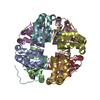
- Structure visualization
| Structure viewer | Molecule: MolmilJmol/JSmol |
|---|
- Downloads & links
-Download
| PDBx/mmCIF format | 2ap6.cif.gz | 176.9 KB | Display | PDBx/mmCIF format |
|---|---|---|---|---|
| PDB format | pdb2ap6.ent.gz | 144 KB | Display | PDB format |
| PDBx/mmJSON format | 2ap6.json.gz | Tree view | PDBx/mmJSON format | |
| Others |  Other downloads Other downloads |
-Validation report
| Arichive directory | https://data.pdbj.org/pub/pdb/validation_reports/ap/2ap6ftp://data.pdbj.org/pub/pdb/validation_reports/ap/2ap6 | HTTPS FTP |
|---|
-Related structure data
| Related structure data |  1vqsS S: Starting model for refinement |
|---|---|
| Similar structure data | |
| Other databases |

-Links
 PDBj
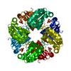
PDBj- Assembly
| Deposited unit | 
| ||||||||
|---|---|---|---|---|---|---|---|---|---|
| 1 |
| ||||||||
| Unit cell |
|
-Components
| #1: Protein | Mass: 13444.924 Da / Num. of mol.: 8 Source method: isolated from a genetically manipulated source Source: (gene. exp.) Agrobacterium tumefaciens str. (bacteria)Species: Agrobacterium tumefaciens / Strain: C58 / Gene: Atu4242 / Production host: #2: Water | ChemComp-HOH / |  Mass: 18.015 Da / Num. of mol.: 184 / Source method: isolated from a natural source / Formula: H2O Mass: 18.015 Da / Num. of mol.: 184 / Source method: isolated from a natural source / Formula: H2OHas protein modification | Y | |
|---|
-Experimental details
-Experiment
| Experiment | Method: X-RAY DIFFRACTION / Number of used crystals: 1 |
|---|
- Sample preparation
| Crystal | Density Matthews: 2.41 Å3/Da / Density % sol: 49.12 % |
|---|
-Data collection
| Diffraction | Mean temperature: 100 K |
|---|---|
| Diffraction source | Source: SYNCHROTRON / Site: NSLS  / Beamline: X4A / Wavelength: 0.97908 / Wavelength: 0.97908 Å / Beamline: X4A / Wavelength: 0.97908 / Wavelength: 0.97908 Å |
| Detector | Type: ADSC QUANTUM 4 / Detector: CCD / Date: Apr 1, 2005 |
| Radiation | Monochromator: SI 111 CHANNEL / Protocol: SINGLE WAVELENGTH / Monochromatic (M) / Laue (L): M / Scattering type: x-ray |
| Radiation wavelength | Wavelength: 0.97908 Å / Relative weight: 1 |
| Reflection | Resolution: 2.5→20 Å / Num. obs: 61871 / % possible obs: 90.3 % / Observed criterion σ(I): -3 / Redundancy: 3.7 % / Biso Wilson estimate: 20.7 Å2 / Rmerge(I) obs: 0.062 / Net I/σ(I): 26.17 |
| Reflection shell | Resolution: 2.5→2.59 Å / Redundancy: 2.6 % / Rmerge(I) obs: 0.182 / Mean I/σ(I) obs: 6.28 / % possible all: 86.4 |
- Processing
| Software |
| ||||||||||||||||||||||||||||||||||||||||||||||||||||||||||||
|---|---|---|---|---|---|---|---|---|---|---|---|---|---|---|---|---|---|---|---|---|---|---|---|---|---|---|---|---|---|---|---|---|---|---|---|---|---|---|---|---|---|---|---|---|---|---|---|---|---|---|---|---|---|---|---|---|---|---|---|---|---|
| Refinement | Method to determine structure: MOLECULAR REPLACEMENT Starting model: PDB Entry 1VQS Resolution: 2.5→19.72 Å / Rfactor Rfree error: 0.007 / Data cutoff high absF: 259473.82 / Data cutoff low absF: 0 / Isotropic thermal model: RESTRAINED / Cross valid method: THROUGHOUT / σ(F): 0 / Stereochemistry target values: Engh & Huber
| ||||||||||||||||||||||||||||||||||||||||||||||||||||||||||||
| Solvent computation | Solvent model: FLAT MODEL / Bsol: 26.6857 Å2 / ksol: 0.390826 e/Å3 | ||||||||||||||||||||||||||||||||||||||||||||||||||||||||||||
| Displacement parameters | Biso mean: 11.9 Å2
| ||||||||||||||||||||||||||||||||||||||||||||||||||||||||||||
| Refine analyze |
| ||||||||||||||||||||||||||||||||||||||||||||||||||||||||||||
| Refinement step | Cycle: LAST / Resolution: 2.5→19.72 Å
| ||||||||||||||||||||||||||||||||||||||||||||||||||||||||||||
| Refine LS restraints |
| ||||||||||||||||||||||||||||||||||||||||||||||||||||||||||||
| Refine LS restraints NCS | NCS model details: CONSTR | ||||||||||||||||||||||||||||||||||||||||||||||||||||||||||||
| LS refinement shell | Resolution: 2.5→2.66 Å / Rfactor Rfree error: 0.027 / Total num. of bins used: 6
| ||||||||||||||||||||||||||||||||||||||||||||||||||||||||||||
| Xplor file |
|
