 Movie
Movie Controller

[English] 日本語
 Yorodumi
- PDB-2aja: X-Ray structure of an ankyrin repeat family protein Q5ZSV0 from L... -
+ Open data

- Basic information
| Entry | Database: PDB / ID: 2aja | ||||||
|---|---|---|---|---|---|---|---|
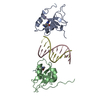
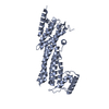
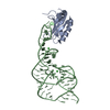
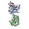
| Title | X-Ray structure of an ankyrin repeat family protein Q5ZSV0 from Legionella pneumophila. Northeast Structural Genomics Consortium target LgR21. | ||||||
 Components Components | ankyrin repeat family protein | ||||||
 Keywords Keywords | PROTEIN BINDING / NESG / Q5ZSV0 / Structural Genomics / PSI / Protein Structure Initiative / Northeast Structural Genomics Consortium | ||||||
| Function / homology | Ankyrin repeat-containing domain / Ankyrin repeat-containing domain superfamily / Serine Threonine Protein Phosphatase 5, Tetratricopeptide repeat / Alpha Horseshoe / Mainly Alpha / Ankyrin repeat family protein Function and homology information Function and homology information | ||||||
| Biological species |   Legionella pneumophila (bacteria) Legionella pneumophila (bacteria) | ||||||
| Method |  X-RAY DIFFRACTION / SYNCHROTRON / SAD / Resolution: 2.8 Å X-RAY DIFFRACTION / SYNCHROTRON / SAD / Resolution: 2.8 Å | ||||||
 Authors Authors | Kuzin, A.P. / Chen, Y. / Acton, T. / Xiao, R. / Conover, K. / Ma, C. / Kellie, R. / Montelione, G.T. / Tong, L. / Hunt, J.F. / Northeast Structural Genomics Consortium (NESG) | ||||||
 Citation Citation | Journal: To be Published Title: X-Ray structure of an ankyrin repeat family protein Q5ZSV0 from Legionella pneumophila. Authors: Kuzin, A.P. / Chen, Y. / Acton, T. / Xiao, R. / Conover, K. / Ma, C. / Kellie, R. / Montelione, G.T. / Tong, L. / Hunt, J.F. | ||||||
| History |
|
- Structure visualization
| Structure viewer | Molecule: MolmilJmol/JSmol |
|---|
- Downloads & links
-Download
| PDBx/mmCIF format | 2aja.cif.gz | 147 KB | Display | PDBx/mmCIF format |
|---|---|---|---|---|
| PDB format | pdb2aja.ent.gz | 116.8 KB | Display | PDB format |
| PDBx/mmJSON format | 2aja.json.gz | Tree view | PDBx/mmJSON format | |
| Others |  Other downloads Other downloads |
-Validation report
| Arichive directory | https://data.pdbj.org/pub/pdb/validation_reports/aj/2ajaftp://data.pdbj.org/pub/pdb/validation_reports/aj/2aja | HTTPS FTP |
|---|
-Related structure data
| Similar structure data | |
|---|---|
| Other databases |


-Links
 PDBj
PDBj- Assembly
| Deposited unit | 
| ||||||||
|---|---|---|---|---|---|---|---|---|---|
| 1 | 
| ||||||||
| 2 | 
| ||||||||
| Unit cell |
|
-Components
| #1: Protein | Mass: 43265.336 Da / Num. of mol.: 2 Source method: isolated from a genetically manipulated source Source: (gene. exp.) Legionella pneumophila (bacteria) / Plasmid: pET21 / Production host: #2: Water | ChemComp-HOH / |  Mass: 18.015 Da / Num. of mol.: 139 / Source method: isolated from a natural source / Formula: H2O Mass: 18.015 Da / Num. of mol.: 139 / Source method: isolated from a natural source / Formula: H2OHas protein modification | Y | |
|---|
-Experimental details
-Experiment
| Experiment | Method: X-RAY DIFFRACTION / Number of used crystals: 1 |
|---|
- Sample preparation
| Crystal | Density Matthews: 2.5 Å3/Da / Density % sol: 51 % |
|---|---|
| Crystal grow | Temperature: 293 K / Method: vapor diffusion, hanging drop / pH: 7 Details: 0.2M potassium thiocyanate, 20% PEG 3350, 20 mM EDTA, pH 7.0, VAPOR DIFFUSION, HANGING DROP, temperature 293K |
-Data collection
| Diffraction | Mean temperature: 100 K |
|---|---|
| Diffraction source | Source: SYNCHROTRON / Site: NSLS  / Beamline: X4A / Wavelength: 0.979 / Wavelength: 0.979 Å / Beamline: X4A / Wavelength: 0.979 / Wavelength: 0.979 Å |
| Detector | Type: ADSC QUANTUM 4 / Detector: CCD / Date: Jul 12, 2005 |
| Radiation | Protocol: SINGLE WAVELENGTH / Monochromatic (M) / Laue (L): M / Scattering type: x-ray |
| Radiation wavelength | Wavelength: 0.979 Å / Relative weight: 1 |
| Reflection | Resolution: 2.8→30 Å / Num. all: 40286 / % possible obs: 99.9 % / Observed criterion σ(I): -3 / Redundancy: 4.95 % / Biso Wilson estimate: 28.2 Å2 / Rsym value: 0.07 |
| Reflection shell | Resolution: 2.8→2.9 Å / Rsym value: 0.235 / % possible all: 99.9 |
- Processing
| Software |
| ||||||||||||||||||||
|---|---|---|---|---|---|---|---|---|---|---|---|---|---|---|---|---|---|---|---|---|---|
| Refinement | Method to determine structure: SAD / Resolution: 2.8→19.92 Å / Rfactor Rfree error: 0.007 / Data cutoff high absF: 287667.08 / Data cutoff low absF: 0 / Isotropic thermal model: RESTRAINED / Cross valid method: THROUGHOUT / σ(F): 2 / Stereochemistry target values: Engh & Huber
| ||||||||||||||||||||
| Solvent computation | Solvent model: FLAT MODEL / Bsol: 13.9618 Å2 / ksol: 0.27268 e/Å3 | ||||||||||||||||||||
| Displacement parameters | Biso mean: 35.5 Å2
| ||||||||||||||||||||
| Refine analyze |
| ||||||||||||||||||||
| Refinement step | Cycle: LAST / Resolution: 2.8→19.92 Å
| ||||||||||||||||||||
| Refine LS restraints |
| ||||||||||||||||||||
| LS refinement shell | Resolution: 2.8→2.97 Å / Rfactor Rfree error: 0.018 / Total num. of bins used: 6
| ||||||||||||||||||||
| Xplor file |
|
