 Movie
Movie Controller

[English] 日本語
 Yorodumi
- PDB-2ahv: Crystal Structure of Acyl-CoA transferase from E. coli O157:H7 (Y... -
+ Open data

- Basic information
| Entry | Database: PDB / ID: 2ahv | ||||||
|---|---|---|---|---|---|---|---|
| Title | Crystal Structure of Acyl-CoA transferase from E. coli O157:H7 (YdiF)-thioester complex with CoA- 1 | ||||||
 Components Components | putative enzyme YdiF | ||||||
 Keywords Keywords | TRANSFERASE / YdiF / CoA transferase / Glutamyl thioester / Structural Genomics / Montreal-Kingston Bacterial Structural Genomics Initiative / BSGI | ||||||
| Function / homology |  Function and homology information Function and homology informationacetate CoA-transferase / Transferases; Transferring sulfur-containing groups; CoA-transferases / ketone body catabolic process / short-chain fatty acid metabolic process / acetate CoA-transferase activity / protein homotetramerization Similarity search - Function | ||||||
| Biological species |  | ||||||
| Method |  X-RAY DIFFRACTION / SYNCHROTRON / MOLECULAR REPLACEMENT / Resolution: 2 Å X-RAY DIFFRACTION / SYNCHROTRON / MOLECULAR REPLACEMENT / Resolution: 2 Å | ||||||
 Authors Authors | Rangarajan, E.S. / Li, Y. / Ajamian, E. / Iannuzzi, P. / Kernaghan, S.D. / Fraser, M.E. / Cygler, M. / Matte, A. / Montreal-Kingston Bacterial Structural Genomics Initiative (BSGI) | ||||||
 Citation Citation | Journal: J.Biol.Chem. / Year: 2005 Title: Crystallographic trapping of the glutamyl-CoA thioester intermediate of family I CoA transferases. Authors: Rangarajan, E.S. / Li, Y. / Ajamian, E. / Iannuzzi, P. / Kernaghan, S.D. / Fraser, M.E. / Cygler, M. / Matte, A. | ||||||
| History |
|
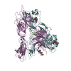
- Structure visualization
| Structure viewer | Molecule: MolmilJmol/JSmol |
|---|
- Downloads & links
-Download
| PDBx/mmCIF format | 2ahv.cif.gz | 423.6 KB | Display | PDBx/mmCIF format |
|---|---|---|---|---|
| PDB format | pdb2ahv.ent.gz | 345.1 KB | Display | PDB format |
| PDBx/mmJSON format | 2ahv.json.gz | Tree view | PDBx/mmJSON format | |
| Others |  Other downloads Other downloads |
-Validation report
| Arichive directory | https://data.pdbj.org/pub/pdb/validation_reports/ah/2ahvftp://data.pdbj.org/pub/pdb/validation_reports/ah/2ahv | HTTPS FTP |
|---|
-Related structure data
| Related structure data | 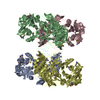 2ahuSC  2ahwC S: Starting model for refinement C: citing same article ( |
|---|---|
| Similar structure data | |
| Other databases |


-Links
 PDBj
PDBj


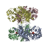
- Assembly
| Deposited unit | 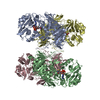
| ||||||||
|---|---|---|---|---|---|---|---|---|---|
| 1 |
| ||||||||
| Unit cell |
|
-Components
| #1: Protein | Mass: 57566.914 Da / Num. of mol.: 4 Source method: isolated from a genetically manipulated source Source: (gene. exp.) References: UniProt: Q8X5X6, Transferases; Transferring sulfur-containing groups; CoA-transferases #2: Chemical | ChemComp-COA /   Mass: 767.534 Da / Num. of mol.: 4 / Source method: obtained synthetically / Formula: C21H36N7O16P3S Mass: 767.534 Da / Num. of mol.: 4 / Source method: obtained synthetically / Formula: C21H36N7O16P3S#3: Water | ChemComp-HOH / |  Mass: 18.015 Da / Num. of mol.: 1474 / Source method: isolated from a natural source / Formula: H2O Mass: 18.015 Da / Num. of mol.: 1474 / Source method: isolated from a natural source / Formula: H2OHas protein modification | Y | |
|---|
-Experimental details
-Experiment
| Experiment | Method: X-RAY DIFFRACTION / Number of used crystals: 1 |
|---|
- Sample preparation
| Crystal | Density Matthews: 2.43 Å3/Da / Density % sol: 48 % |
|---|---|
| Crystal grow | Temperature: 293 K / Method: microbatch / pH: 7.5 Details: 16% (w/v) PEG 3350, 80 mM Na K tartarate, pH 7.5, Microbatch, temperature 293K |
-Data collection
| Diffraction | Mean temperature: 100 K |
|---|---|
| Diffraction source | Source: SYNCHROTRON / Site: NSLS  / Beamline: X29A / Wavelength: 1.1 Å / Beamline: X29A / Wavelength: 1.1 Å |
| Detector | Type: ADSC QUANTUM 315 / Detector: CCD / Date: Mar 25, 2005 |
| Radiation | Monochromator: silicone / Protocol: SINGLE WAVELENGTH / Monochromatic (M) / Laue (L): M / Scattering type: x-ray |
| Radiation wavelength | Wavelength: 1.1 Å / Relative weight: 1 |
| Reflection | Resolution: 2→50 Å / Num. all: 159093 / Num. obs: 159093 / % possible obs: 98.8 % / Observed criterion σ(F): 0 / Observed criterion σ(I): 0 / Redundancy: 3.6 % / Biso Wilson estimate: 25.8 Å2 / Rsym value: 0.079 / Net I/σ(I): 9 |
| Reflection shell | Resolution: 2→2.07 Å / Redundancy: 2.7 % / Mean I/σ(I) obs: 2 / Num. unique all: 14536 / Rsym value: 0.454 / % possible all: 90.5 |
- Processing
| Software |
| ||||||||||||||||||||||||||||||||||||||||||||||||||||||||||||||||||||||||||||||||||||||||||
|---|---|---|---|---|---|---|---|---|---|---|---|---|---|---|---|---|---|---|---|---|---|---|---|---|---|---|---|---|---|---|---|---|---|---|---|---|---|---|---|---|---|---|---|---|---|---|---|---|---|---|---|---|---|---|---|---|---|---|---|---|---|---|---|---|---|---|---|---|---|---|---|---|---|---|---|---|---|---|---|---|---|---|---|---|---|---|---|---|---|---|---|
| Refinement | Method to determine structure: MOLECULAR REPLACEMENT Starting model: PDB Entry 2AHU Resolution: 2→50 Å / Cor.coef. Fo:Fc: 0.955 / Cor.coef. Fo:Fc free: 0.932 / SU B: 4.028 / SU ML: 0.112 / Cross valid method: THROUGHOUT / σ(F): 0 / ESU R: 0.185 / ESU R Free: 0.163 / Stereochemistry target values: MAXIMUM LIKELIHOOD
| ||||||||||||||||||||||||||||||||||||||||||||||||||||||||||||||||||||||||||||||||||||||||||
| Solvent computation | Ion probe radii: 0.8 Å / Shrinkage radii: 0.8 Å / VDW probe radii: 1.2 Å / Solvent model: BABINET MODEL WITH MASK | ||||||||||||||||||||||||||||||||||||||||||||||||||||||||||||||||||||||||||||||||||||||||||
| Displacement parameters | Biso mean: 25.933 Å2
| ||||||||||||||||||||||||||||||||||||||||||||||||||||||||||||||||||||||||||||||||||||||||||
| Refinement step | Cycle: LAST / Resolution: 2→50 Å
| ||||||||||||||||||||||||||||||||||||||||||||||||||||||||||||||||||||||||||||||||||||||||||
| Refine LS restraints |
| ||||||||||||||||||||||||||||||||||||||||||||||||||||||||||||||||||||||||||||||||||||||||||
| LS refinement shell | Resolution: 2.001→2.053 Å / Total num. of bins used: 20
|
