 Movie
Movie Controller

[English] 日本語
 Yorodumi
- PDB-2agx: Crystal structure of the Schiff base intermediate in the reductiv... -
+ Open data

- Basic information
| Entry | Database: PDB / ID: 2agx | ||||||
|---|---|---|---|---|---|---|---|
| Title | Crystal structure of the Schiff base intermediate in the reductive half-reaction of aromatic amine dehydrogenase (AADH) with tryptamine. P212121 form | ||||||
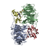
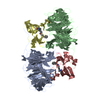
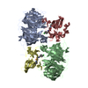
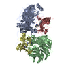
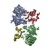
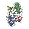
 Components Components | (Aromatic amine dehydrogenase) x 2 | ||||||
 Keywords Keywords | OXIDOREDUCTASE | ||||||
| Function / homology |  Function and homology information Function and homology informationaralkylamine dehydrogenase (azurin) / aralkylamine dehydrogenase (azurin) activity / aliphatic amine dehydrogenase activity / amine metabolic process / periplasmic space Similarity search - Function | ||||||
| Biological species |  Alcaligenes faecalis (bacteria) Alcaligenes faecalis (bacteria) | ||||||
| Method |  X-RAY DIFFRACTION / SYNCHROTRON / MOLECULAR REPLACEMENT / Resolution: 2.2 Å X-RAY DIFFRACTION / SYNCHROTRON / MOLECULAR REPLACEMENT / Resolution: 2.2 Å | ||||||
 Authors Authors | Masgrau, L. / Roujeinikova, A. / Johannissen, L.O. / Hothi, P. / Basran, J. / Ranaghan, K.E. / Mulholland, A.J. / Sutcliffe, M.J. / Scrutton, N.S. / Leys, D. | ||||||
 Citation Citation | Journal: Science / Year: 2006 Title: Atomic description of an enzyme reaction dominated by proton tunneling Authors: Masgrau, L. / Roujeinikova, A. / Johannissen, L.O. / Hothi, P. / Basran, J. / Ranaghan, K.E. / Mulholland, A.J. / Sutcliffe, M.J. / Scrutton, N.S. / Leys, D. | ||||||
| History |
|
- Structure visualization
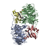
| Structure viewer | Molecule: MolmilJmol/JSmol |
|---|
- Downloads & links
-Download
| PDBx/mmCIF format | 2agx.cif.gz | 206.6 KB | Display | PDBx/mmCIF format |
|---|---|---|---|---|
| PDB format | pdb2agx.ent.gz | 162.2 KB | Display | PDB format |
| PDBx/mmJSON format | 2agx.json.gz | Tree view | PDBx/mmJSON format | |
| Others |  Other downloads Other downloads |
-Validation report
| Arichive directory | https://data.pdbj.org/pub/pdb/validation_reports/ag/2agxftp://data.pdbj.org/pub/pdb/validation_reports/ag/2agx | HTTPS FTP |
|---|
-Related structure data
| Related structure data | 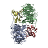 2aglC  2agwC  2agyC  2agzC 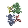 2ah0C  2ah1C  2agu C: citing same article ( |
|---|---|
| Similar structure data |


-Links
 PDBj
PDBj

- Assembly
| Deposited unit | 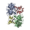
| ||||||||
|---|---|---|---|---|---|---|---|---|---|
| 1 |
| ||||||||
| Unit cell |
| ||||||||
| Details | The asymmetric unit contains the biological unit (heterotetramer) |
-Components
| #1: Protein | Mass: 14516.898 Da / Num. of mol.: 2 / Fragment: residues 48-182 / Source method: isolated from a natural source / Source: (natural) Alcaligenes faecalis (bacteria) / Strain: IFO 14479 / References: UniProt: P84887, EC: 1.4.99.4#2: Protein | Mass: 40016.125 Da / Num. of mol.: 2 / Fragment: residues 73-433 / Source method: isolated from a natural source / Source: (natural) Alcaligenes faecalis (bacteria) / Strain: IFO 14479 / References: UniProt: P84888, EC: 1.4.99.4#3: Chemical | 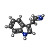  Mass: 158.200 Da / Num. of mol.: 2 / Source method: obtained synthetically / Formula: C10H10N2 Mass: 158.200 Da / Num. of mol.: 2 / Source method: obtained synthetically / Formula: C10H10N2#4: Water | ChemComp-HOH / |  Mass: 18.015 Da / Num. of mol.: 577 / Source method: isolated from a natural source / Formula: H2O Mass: 18.015 Da / Num. of mol.: 577 / Source method: isolated from a natural source / Formula: H2OHas protein modification | Y | |
|---|
-Experimental details
-Experiment
| Experiment | Method: X-RAY DIFFRACTION / Number of used crystals: 1 |
|---|
- Sample preparation
| Crystal | Density Matthews: 2.4 Å3/Da / Density % sol: 48.7 % |
|---|---|
| Crystal grow | Temperature: 292 K / Method: vapor diffusion, sitting drop / pH: 6 Details: PEG 2000 MME, ammonium sulphate, sodium cacodylate, tryptamine, pH 6.0, VAPOR DIFFUSION, SITTING DROP, temperature 292K |
-Data collection
| Diffraction | Mean temperature: 100 K |
|---|---|
| Diffraction source | Source: SYNCHROTRON / Site: EMBL/DESY, HAMBURG  / Beamline: X11 / Wavelength: 0.8 Å / Beamline: X11 / Wavelength: 0.8 Å |
| Detector | Type: MARRESEARCH / Detector: CCD |
| Radiation | Protocol: SINGLE WAVELENGTH / Monochromatic (M) / Laue (L): M / Scattering type: x-ray |
| Radiation wavelength | Wavelength: 0.8 Å / Relative weight: 1 |
| Reflection | Resolution: 2.2→30 Å / Num. obs: 49153 / % possible obs: 91 % / Observed criterion σ(I): -3 / Redundancy: 4.7 % / Biso Wilson estimate: 25.3 Å2 / Rmerge(I) obs: 0.1 / Net I/σ(I): 12.6 |
| Reflection shell | Resolution: 2.2→2.28 Å / Rmerge(I) obs: 0.332 / Mean I/σ(I) obs: 28 / % possible all: 75 |
- Processing
| Software |
| |||||||||||||||||||||||||||||||||||||||||||||||||||||||||||||||||||||||||||||||||||||
|---|---|---|---|---|---|---|---|---|---|---|---|---|---|---|---|---|---|---|---|---|---|---|---|---|---|---|---|---|---|---|---|---|---|---|---|---|---|---|---|---|---|---|---|---|---|---|---|---|---|---|---|---|---|---|---|---|---|---|---|---|---|---|---|---|---|---|---|---|---|---|---|---|---|---|---|---|---|---|---|---|---|---|---|---|---|---|
| Refinement | Method to determine structure: MOLECULAR REPLACEMENT Starting model: substrate-free AADH coordinates Resolution: 2.2→15 Å / Cor.coef. Fo:Fc: 0.957 / Cor.coef. Fo:Fc free: 0.917 / SU B: 5.26 / SU ML: 0.134 / Cross valid method: THROUGHOUT / σ(F): 0 / ESU R: 0.268 / ESU R Free: 0.214 / Stereochemistry target values: MAXIMUM LIKELIHOOD
| |||||||||||||||||||||||||||||||||||||||||||||||||||||||||||||||||||||||||||||||||||||
| Solvent computation | Ion probe radii: 0.8 Å / Shrinkage radii: 0.8 Å / VDW probe radii: 1.2 Å / Solvent model: BABINET MODEL WITH MASK | |||||||||||||||||||||||||||||||||||||||||||||||||||||||||||||||||||||||||||||||||||||
| Displacement parameters | Biso mean: 22.518 Å2
| |||||||||||||||||||||||||||||||||||||||||||||||||||||||||||||||||||||||||||||||||||||
| Refinement step | Cycle: LAST / Resolution: 2.2→15 Å
| |||||||||||||||||||||||||||||||||||||||||||||||||||||||||||||||||||||||||||||||||||||
| Refine LS restraints |
| |||||||||||||||||||||||||||||||||||||||||||||||||||||||||||||||||||||||||||||||||||||
| LS refinement shell | Resolution: 2.2→2.256 Å / Total num. of bins used: 20
|
