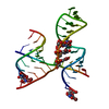
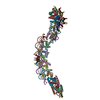
ジャーナル: Structure / 年: 2005 タイトル: Structure of the hepatitis C virus IRES bound to the human 80S ribosome: remodeling of the HCV IRES. 著者: Daniel Boehringer / Rolf Thermann / Antje Ostareck-Lederer / Joe D Lewis / Holger Stark / 要旨: Initiation of translation of the hepatitis C virus (HCV) polyprotein is driven by an internal ribosome entry site (IRES) RNA that bypasses much of the eukaryotic translation initiation machinery. ...Initiation of translation of the hepatitis C virus (HCV) polyprotein is driven by an internal ribosome entry site (IRES) RNA that bypasses much of the eukaryotic translation initiation machinery. Here, single-particle electron cryomicroscopy has been used to study the mechanism of HCV IRES-mediated initiation. A HeLa in vitro translation system was used to assemble human IRES-80S ribosome complexes under near physiological conditions; these were stalled before elongation. Domain 2 of the HCV IRES is bound to the tRNA exit site, touching the L1 stalk of the 60S subunit, suggesting a mechanism for the removal of the HCV IRES in the progression to elongation. Domain 3 of the HCV IRES positions the initiation codon in the ribosomal mRNA binding cleft by binding helix 28 at the head of the 40S subunit. The comparison with the previously published binary 40S-HCV IRES complex reveals structural rearrangements in the two pseudoknot structures of the HCV IRES in translation initiation.
プロトコル: SINGLE WAVELENGTH / 単色(M)・ラウエ(L): M / 散乱光タイプ: x-ray
放射波長
相対比: 1
-
解析
EMソフトウェア
ID
名称
バージョン
カテゴリ
1
IMAGIC
3次元再構成
2
Amira
2.3
モデルフィッティング
CTF補正
詳細: phase reversal
対称性
点対称性: C1 (非対称)
3次元再構成
手法: angular reconstitution / 解像度: 15 Å / 粒子像の数: 24100 / ピクセルサイズ(公称値): 3.6 Å / ピクセルサイズ(実測値): 3.6 Å 詳細: This entry contains only phosphorus atom in the coordinate. 対称性のタイプ: POINT
 ムービー
ムービー コントローラー


 データを開く
 基本情報
基本情報 要素
要素 キーワード
キーワード 機能・相同性情報
 データ登録者
 引用
引用
 構造の表示
構造の表示 ダウンロードとリンク
 その他のダウンロード
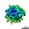
 PDBj
PDBj


 集合体
集合体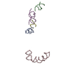
 試料調製
試料調製 電子顕微鏡撮影
 FIELD EMISSION GUN / 加速電圧: 200 kV / 照射モード: FLOOD BEAM
 解析
解析