 Movie
Movie Controller


+ Open data

- Basic information
| Entry | Database: PDB / ID: 2ae2 | ||||||
|---|---|---|---|---|---|---|---|
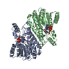
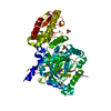
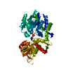
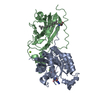
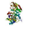
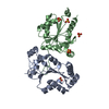
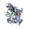
| Title | TROPINONE REDUCTASE-II COMPLEXED WITH NADP+ AND PSEUDOTROPINE | ||||||
 Components Components | PROTEIN (TROPINONE REDUCTASE-II) | ||||||
 Keywords Keywords | OXIDOREDUCTASE / TROPANE ALKALOID BIOSYNTHESIS / REDUCTION OF TROPINONE TO PSEUDOTROPINE / SHORT-CHAIN DEHYDROGENASE | ||||||
| Function / homology |  Function and homology information Function and homology informationtropinone reductase II / tropinone reductase activity / tropane alkaloid biosynthetic process Similarity search - Function | ||||||
| Biological species |  Datura stramonium (jimsonweed) Datura stramonium (jimsonweed) | ||||||
| Method |  X-RAY DIFFRACTION / SYNCHROTRON / MOLECULAR REPLACEMENT / Resolution: 1.9 Å X-RAY DIFFRACTION / SYNCHROTRON / MOLECULAR REPLACEMENT / Resolution: 1.9 Å | ||||||
 Authors Authors | Yamashita, A. / Kato, H. / Wakatsuki, S. / Tomizaki, T. / Nakatsu, T. / Nakajima, K. / Hashimoto, T. / Yamada, Y. / Oda, J. | ||||||
 Citation Citation | Journal: Biochemistry / Year: 1999 Title: Structure of tropinone reductase-II complexed with NADP+ and pseudotropine at 1.9 A resolution: implication for stereospecific substrate binding and catalysis. Authors: Yamashita, A. / Kato, H. / Wakatsuki, S. / Tomizaki, T. / Nakatsu, T. / Nakajima, K. / Hashimoto, T. / Yamada, Y. / Oda, J. #1: Journal: Proc.Natl.Acad.Sci.USA / Year: 1998Title: Crystal structures of two tropinone reductases: different reaction stereospecificities in the same protein fold. Authors: Nakajima, K. / Yamashita, A. / Akama, H. / Nakatsu, T. / Kato, H. / Hashimoto, T. / Oda, J. / Yamada, Y. | ||||||
| History |
|
- Structure visualization
| Structure viewer | Molecule: MolmilJmol/JSmol |
|---|
- Downloads & links
-Download
| PDBx/mmCIF format | 2ae2.cif.gz | 118.2 KB | Display | PDBx/mmCIF format |
|---|---|---|---|---|
| PDB format | pdb2ae2.ent.gz | 91.6 KB | Display | PDB format |
| PDBx/mmJSON format | 2ae2.json.gz | Tree view | PDBx/mmJSON format | |
| Others |  Other downloads Other downloads |
-Validation report
| Arichive directory | https://data.pdbj.org/pub/pdb/validation_reports/ae/2ae2ftp://data.pdbj.org/pub/pdb/validation_reports/ae/2ae2 | HTTPS FTP |
|---|
-Related structure data
| Related structure data | 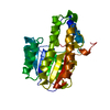 2ae1S S: Starting model for refinement |
|---|---|
| Similar structure data |


-Links
 PDBj
PDBj


- Assembly
| Deposited unit | 
| ||||||||
|---|---|---|---|---|---|---|---|---|---|
| 1 |
| ||||||||
| 2 | 
| ||||||||
| 3 | 
| ||||||||
| Unit cell |
| ||||||||
| Noncrystallographic symmetry (NCS) | NCS oper: (Code: given Matrix: (-0.89137, 0.19074, -0.41118), Vector: |
-Components
| #1: Protein | Mass: 28339.445 Da / Num. of mol.: 2 Source method: isolated from a genetically manipulated source Source: (gene. exp.) Datura stramonium (jimsonweed) / Organ: CULTURED ROOT / Plasmid: PETTR2 / Species (production host): Escherichia coli / Production host:  #2: Chemical |   Mass: 743.405 Da / Num. of mol.: 2 / Source method: obtained synthetically / Formula: C21H28N7O17P3 Mass: 743.405 Da / Num. of mol.: 2 / Source method: obtained synthetically / Formula: C21H28N7O17P3#3: Chemical |   Mass: 141.211 Da / Num. of mol.: 2 / Source method: obtained synthetically / Formula: C8H15NO Mass: 141.211 Da / Num. of mol.: 2 / Source method: obtained synthetically / Formula: C8H15NO#4: Water | ChemComp-HOH / |  Mass: 18.015 Da / Num. of mol.: 236 / Source method: isolated from a natural source / Formula: H2O Mass: 18.015 Da / Num. of mol.: 236 / Source method: isolated from a natural source / Formula: H2O |
|---|
-Experimental details
-Experiment
| Experiment | Method: X-RAY DIFFRACTION / Number of used crystals: 1 |
|---|
- Sample preparation
| Crystal | Density Matthews: 3.42 Å3/Da / Density % sol: 64.1 % | ||||||||||||||||||||||||||||||||||||||||||||||||||||||
|---|---|---|---|---|---|---|---|---|---|---|---|---|---|---|---|---|---|---|---|---|---|---|---|---|---|---|---|---|---|---|---|---|---|---|---|---|---|---|---|---|---|---|---|---|---|---|---|---|---|---|---|---|---|---|---|
| Crystal grow | pH: 7.5 / Details: pH 7.5 | ||||||||||||||||||||||||||||||||||||||||||||||||||||||
| Crystal grow | *PLUS Temperature: 20 ℃ / Method: vapor diffusion, hanging dropDetails: drop consists of equal volume of protein and reservoir solutions | ||||||||||||||||||||||||||||||||||||||||||||||||||||||
| Components of the solutions | *PLUS
|
-Data collection
| Diffraction | Mean temperature: 293 K |
|---|---|
| Diffraction source | Source: SYNCHROTRON / Site: ESRF  / Beamline: ID14-3 / Wavelength: 0.918 / Beamline: ID14-3 / Wavelength: 0.918 |
| Detector | Detector: IMAGE PLATE / Date: Jul 13, 1997 |
| Radiation | Protocol: SINGLE WAVELENGTH / Monochromatic (M) / Laue (L): M / Scattering type: x-ray |
| Radiation wavelength | Wavelength: 0.918 Å / Relative weight: 1 |
| Reflection | Resolution: 1.9→64.2 Å / Num. obs: 54120 / % possible obs: 85.6 % / Observed criterion σ(I): 1 / Redundancy: 3.6 % / Biso Wilson estimate: 16.9 Å2 / Rmerge(I) obs: 0.084 |
| Reflection shell | Resolution: 1.9→1.93 Å / Rmerge(I) obs: 0.247 / % possible all: 77.9 |
| Reflection | *PLUS Num. measured all: 195899 |
| Reflection shell | *PLUS % possible obs: 77.9 % |
- Processing
| Software |
| ||||||||||||||||||||||||||||||||||||||||||||||||||||||||||||
|---|---|---|---|---|---|---|---|---|---|---|---|---|---|---|---|---|---|---|---|---|---|---|---|---|---|---|---|---|---|---|---|---|---|---|---|---|---|---|---|---|---|---|---|---|---|---|---|---|---|---|---|---|---|---|---|---|---|---|---|---|---|
| Refinement | Method to determine structure: MOLECULAR REPLACEMENT Starting model: PDB ENTRY 2AE1 Resolution: 1.9→10 Å / Cross valid method: THROUGHOUT / σ(F): 2
| ||||||||||||||||||||||||||||||||||||||||||||||||||||||||||||
| Displacement parameters | Biso mean: 19.1 Å2 | ||||||||||||||||||||||||||||||||||||||||||||||||||||||||||||
| Refine analyze |
| ||||||||||||||||||||||||||||||||||||||||||||||||||||||||||||
| Refinement step | Cycle: LAST / Resolution: 1.9→10 Å
| ||||||||||||||||||||||||||||||||||||||||||||||||||||||||||||
| Refine LS restraints |
| ||||||||||||||||||||||||||||||||||||||||||||||||||||||||||||
| Refine LS restraints NCS | NCS model details: UNRESTRAINED | ||||||||||||||||||||||||||||||||||||||||||||||||||||||||||||
| LS refinement shell | Resolution: 1.9→1.99 Å / Total num. of bins used: 8
| ||||||||||||||||||||||||||||||||||||||||||||||||||||||||||||
| Xplor file |
| ||||||||||||||||||||||||||||||||||||||||||||||||||||||||||||
| Software | *PLUS Name: X-PLOR / Version: 3.851 / Classification: refinement | ||||||||||||||||||||||||||||||||||||||||||||||||||||||||||||
| Refine LS restraints | *PLUS
|
