 Movie
Movie Controller

[English] 日本語
 Yorodumi
- PDB-2aag: Crystal Structures of the Wild-type, Mutant-P1A and Inactivated M... -
+ Open data

- Basic information
| Entry | Database: PDB / ID: 2aag | ||||||
|---|---|---|---|---|---|---|---|
| Title | Crystal Structures of the Wild-type, Mutant-P1A and Inactivated Malonate Semialdehyde Decarboxylase: A Structural Basis for the Decarboxylase and Hydratase Activities | ||||||
 Components Components | Malonate Semialdehyde Decarboxylase | ||||||
 Keywords Keywords | LYASE / tautomerase superfamily / beta-alpha-beta / homotrimeric | ||||||
| Function / homology | Tautomerase, MSAD family / Tautomerase enzyme / Macrophage Migration Inhibitory Factor / Macrophage Migration Inhibitory Factor / Tautomerase/MIF superfamily / 2-Layer Sandwich / Alpha Beta / Malonate semialdehyde decarboxylase Function and homology information Function and homology information | ||||||
| Biological species |  Pseudomonas pavonaceae (bacteria) Pseudomonas pavonaceae (bacteria) | ||||||
| Method |  X-RAY DIFFRACTION / MOLECULAR REPLACEMENT / Resolution: 1.85 Å X-RAY DIFFRACTION / MOLECULAR REPLACEMENT / Resolution: 1.85 Å | ||||||
 Authors Authors | Almrud, J.J. / Poelarends, G.J. / Johnson Jr., W.H. / Serrano, H. / Hackert, M.L. / Whitman, C.P. | ||||||
 Citation Citation | Journal: Biochemistry / Year: 2005 Title: Crystal Structures of the Wild-Type, P1A Mutant, and Inactivated Malonate Semialdehyde Decarboxylase: A Structural Basis for the Decarboxylase and Hydratase Activities Authors: Almrud, J.J. / Poelarends, G.J. / Johnson Jr., W.H. / Serrano, H. / Hackert, M.L. / Whitman, C.P. | ||||||
| History |
|
- Structure visualization
| Structure viewer | Molecule: MolmilJmol/JSmol |
|---|
- Downloads & links
-Download
| PDBx/mmCIF format | 2aag.cif.gz | 319.7 KB | Display | PDBx/mmCIF format |
|---|---|---|---|---|
| PDB format | pdb2aag.ent.gz | 261.4 KB | Display | PDB format |
| PDBx/mmJSON format | 2aag.json.gz | Tree view | PDBx/mmJSON format | |
| Others |  Other downloads Other downloads |
-Validation report
| Arichive directory | https://data.pdbj.org/pub/pdb/validation_reports/aa/2aagftp://data.pdbj.org/pub/pdb/validation_reports/aa/2aag | HTTPS FTP |
|---|
-Related structure data


-Links
 PDBj
PDBj- Assembly
| Deposited unit | 
| ||||||||||||||||||||||||||||||||||||||||||||||||||||||||||||||||||||||
|---|---|---|---|---|---|---|---|---|---|---|---|---|---|---|---|---|---|---|---|---|---|---|---|---|---|---|---|---|---|---|---|---|---|---|---|---|---|---|---|---|---|---|---|---|---|---|---|---|---|---|---|---|---|---|---|---|---|---|---|---|---|---|---|---|---|---|---|---|---|---|---|
| 1 | 
| ||||||||||||||||||||||||||||||||||||||||||||||||||||||||||||||||||||||
| 2 | 
| ||||||||||||||||||||||||||||||||||||||||||||||||||||||||||||||||||||||
| Unit cell |
| ||||||||||||||||||||||||||||||||||||||||||||||||||||||||||||||||||||||
| Noncrystallographic symmetry (NCS) | NCS domain:
NCS domain segments: Component-ID: 1 / Ens-ID: 1 / Beg auth comp-ID: PRO / Beg label comp-ID: PRO / Refine code: 1
| ||||||||||||||||||||||||||||||||||||||||||||||||||||||||||||||||||||||
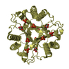
| Details | the biological unit is a homotrimer; two of which crystallized in the asymmetric unit. |
-Components
| #1: Protein | Mass: 14180.961 Da / Num. of mol.: 6 Source method: isolated from a genetically manipulated source Source: (gene. exp.) Pseudomonas pavonaceae (bacteria) / Strain: 170 / Gene: orf130 / Plasmid: pET3a / Species (production host): Escherichia coli / Production host: References: UniProt: Q9EV83, Lyases; Carbon-carbon lyases; Carboxy-lyases #2: Water | ChemComp-HOH / |  Mass: 18.015 Da / Num. of mol.: 807 / Source method: isolated from a natural source / Formula: H2O Mass: 18.015 Da / Num. of mol.: 807 / Source method: isolated from a natural source / Formula: H2O |
|---|
-Experimental details
-Experiment
| Experiment | Method: X-RAY DIFFRACTION / Number of used crystals: 1 |
|---|
- Sample preparation
| Crystal | Density Matthews: 2.1 Å3/Da / Density % sol: 41.5 % |
|---|---|
| Crystal grow | Temperature: 298 K / Method: vapor diffusion, sitting drop / pH: 8 Details: 35% (v/v) 1,6-hexanediol, 200 mM MgCl2, and 100 mM Tris-Cl buffer, pH 8.0, VAPOR DIFFUSION, SITTING DROP, temperature 298K |
-Data collection
| Diffraction | Mean temperature: 100 K |
|---|---|
| Diffraction source | Source: ROTATING ANODE / Type: RIGAKU RUH3R / Wavelength: 1.5418 Å |
| Detector | Type: RIGAKU RAXIS IV / Detector: IMAGE PLATE / Date: Jan 6, 2004 / Details: Osmic Blue Max-Flux Confocal optical system |
| Radiation | Protocol: SINGLE WAVELENGTH / Monochromatic (M) / Laue (L): M / Scattering type: x-ray |
| Radiation wavelength | Wavelength: 1.5418 Å / Relative weight: 1 |
| Reflection | Resolution: 1.83→30 Å / Num. all: 63310 / Num. obs: 60986 / % possible obs: 96.4 % / Observed criterion σ(I): 2 / Redundancy: 8.4 % / Biso Wilson estimate: 22.45 Å2 / Rsym value: 0.072 / Net I/σ(I): 12.2 |
- Processing
| Software |
| |||||||||||||||||||||||||||||||||||||||||||||||||||||||||||||||||||||||||||||||||||||
|---|---|---|---|---|---|---|---|---|---|---|---|---|---|---|---|---|---|---|---|---|---|---|---|---|---|---|---|---|---|---|---|---|---|---|---|---|---|---|---|---|---|---|---|---|---|---|---|---|---|---|---|---|---|---|---|---|---|---|---|---|---|---|---|---|---|---|---|---|---|---|---|---|---|---|---|---|---|---|---|---|---|---|---|---|---|---|
| Refinement | Method to determine structure: MOLECULAR REPLACEMENT Starting model: c-alpha trace of density map resulting from a single-wavelength anomalous difference data set (using Hg's anomalous signal) Resolution: 1.85→19.21 Å / Cor.coef. Fo:Fc: 0.956 / Cor.coef. Fo:Fc free: 0.933 / SU B: 3.351 / SU ML: 0.103 / Isotropic thermal model: anisotropic / Cross valid method: THROUGHOUT / σ(F): 0 / ESU R Free: 0.154 / Stereochemistry target values: MAXIMUM LIKELIHOOD
| |||||||||||||||||||||||||||||||||||||||||||||||||||||||||||||||||||||||||||||||||||||
| Solvent computation | Ion probe radii: 0.8 Å / Shrinkage radii: 0.8 Å / VDW probe radii: 1.4 Å / Solvent model: BABINET MODEL WITH MASK | |||||||||||||||||||||||||||||||||||||||||||||||||||||||||||||||||||||||||||||||||||||
| Displacement parameters | Biso mean: 23.573 Å2
| |||||||||||||||||||||||||||||||||||||||||||||||||||||||||||||||||||||||||||||||||||||
| Refinement step | Cycle: LAST / Resolution: 1.85→19.21 Å
| |||||||||||||||||||||||||||||||||||||||||||||||||||||||||||||||||||||||||||||||||||||
| Refine LS restraints |
| |||||||||||||||||||||||||||||||||||||||||||||||||||||||||||||||||||||||||||||||||||||
| Refine LS restraints NCS | Ens-ID: 1 / Number: 968 / Refine-ID: X-RAY DIFFRACTION
| |||||||||||||||||||||||||||||||||||||||||||||||||||||||||||||||||||||||||||||||||||||
| LS refinement shell | Resolution: 1.85→1.87 Å / Rfactor Rfree error: 0 / Total num. of bins used: 40
|
