 Movie
Movie Controller


+ Open data

- Basic information
| Entry | Database: PDB / ID: 2a9g | ||||||
|---|---|---|---|---|---|---|---|
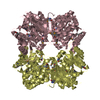
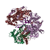
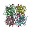
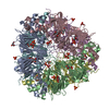
| Title | Structure of C406A arginine deiminase in complex with L-arginine | ||||||
 Components Components | Arginine deiminase | ||||||
 Keywords Keywords | HYDROLASE / arginine degradation pathway / L-arginine deiminase / catalytic mechanism | ||||||
| Function / homology |  Function and homology information Function and homology information | ||||||
| Biological species |   Pseudomonas aeruginosa (bacteria) Pseudomonas aeruginosa (bacteria) | ||||||
| Method |  X-RAY DIFFRACTION / MOLECULAR REPLACEMENT / Resolution: 2.3 Å X-RAY DIFFRACTION / MOLECULAR REPLACEMENT / Resolution: 2.3 Å | ||||||
 Authors Authors | Galkin, A. / Lu, X. / Dunaway-Mariano, D. / Herzberg, O. | ||||||
 Citation Citation | Journal: J.Biol.Chem. / Year: 2005 Title: Crystal Structures Representing the Michaelis Complex and the Thiouronium Reaction Intermediate of Pseudomonas aeruginosa Arginine Deiminase. Authors: Galkin, A. / Lu, X. / Dunaway-Mariano, D. / Herzberg, O. | ||||||
| History |
|
- Structure visualization
| Structure viewer | Molecule: MolmilJmol/JSmol |
|---|
- Downloads & links
-Download
| PDBx/mmCIF format | 2a9g.cif.gz | 343.7 KB | Display | PDBx/mmCIF format |
|---|---|---|---|---|
| PDB format | pdb2a9g.ent.gz | 279 KB | Display | PDB format |
| PDBx/mmJSON format | 2a9g.json.gz | Tree view | PDBx/mmJSON format | |
| Others |  Other downloads Other downloads |
-Validation report
| Arichive directory | https://data.pdbj.org/pub/pdb/validation_reports/a9/2a9gftp://data.pdbj.org/pub/pdb/validation_reports/a9/2a9g | HTTPS FTP |
|---|
-Related structure data
| Related structure data | 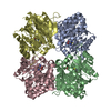 2aafC 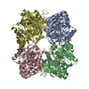 2abrC 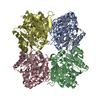 2aciC 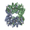 1rxxS S: Starting model for refinement C: citing same article ( |
|---|---|
| Similar structure data |


-Links
 PDBj
PDBj
- Assembly
| Deposited unit | 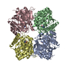
| ||||||||
|---|---|---|---|---|---|---|---|---|---|
| 1 |
| ||||||||
| 2 | 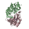
| ||||||||
| 3 | 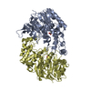
| ||||||||
| Unit cell |
| ||||||||
| Details | The biological assembly is a tetramer generated from the molecules A, B, C and D in the asymmetric unit |
-Components
| #1: Protein | Mass: 46458.824 Da / Num. of mol.: 4 / Mutation: C406A Source method: isolated from a genetically manipulated source Source: (gene. exp.) Pseudomonas aeruginosa (bacteria) / Gene: arcA / Plasmid: PET100-ADIN / Production host: #2: Chemical | ChemComp-ARG /   Type: L-peptide linking / Mass: 175.209 Da / Num. of mol.: 4 / Source method: obtained synthetically / Formula: C6H15N4O2 Type: L-peptide linking / Mass: 175.209 Da / Num. of mol.: 4 / Source method: obtained synthetically / Formula: C6H15N4O2#3: Water | ChemComp-HOH / |  Mass: 18.015 Da / Num. of mol.: 1064 / Source method: isolated from a natural source / Formula: H2O Mass: 18.015 Da / Num. of mol.: 1064 / Source method: isolated from a natural source / Formula: H2O |
|---|
-Experimental details
-Experiment
| Experiment | Method: X-RAY DIFFRACTION / Number of used crystals: 1 |
|---|
- Sample preparation
| Crystal | Density Matthews: 2.23 Å3/Da / Density % sol: 45 % |
|---|---|
| Crystal grow | Temperature: 298 K / Method: vapor diffusion, hanging drop / pH: 7.6 Details: 35% MPD (2-methyl-2,4-pentanediol), 6% PEG 3350, 0.1 M Tris-HCl, L_arginine 0.02 M, pH 7.6, VAPOR DIFFUSION, HANGING DROP, temperature 298.0K |
-Data collection
| Diffraction | Mean temperature: 100 K |
|---|---|
| Diffraction source | Source: ROTATING ANODE / Type: RIGAKU / Wavelength: 1.54178 Å |
| Detector | Type: RIGAKU RAXIS IV / Detector: IMAGE PLATE / Date: Nov 21, 2003 |
| Radiation | Monochromator: Osmic mirrors / Protocol: SINGLE WAVELENGTH / Monochromatic (M) / Laue (L): M / Scattering type: x-ray |
| Radiation wavelength | Wavelength: 1.54178 Å / Relative weight: 1 |
| Reflection | Resolution: 2.3→20 Å / Num. all: 74422 / Num. obs: 73975 / % possible obs: 99.4 % / Rmerge(I) obs: 0.097 |
| Reflection shell | Resolution: 2.3→2.38 Å / Rmerge(I) obs: 0.27 / % possible all: 96.1 |
- Processing
| Software |
| ||||||||||||||||||||
|---|---|---|---|---|---|---|---|---|---|---|---|---|---|---|---|---|---|---|---|---|---|
| Refinement | Method to determine structure: MOLECULAR REPLACEMENT Starting model: 1RXX Resolution: 2.3→20 Å / Cross valid method: THROUGHOUT / σ(F): 0 / Stereochemistry target values: Engh & Huber
| ||||||||||||||||||||
| Displacement parameters | Biso mean: 37 Å2 | ||||||||||||||||||||
| Refinement step | Cycle: LAST / Resolution: 2.3→20 Å
| ||||||||||||||||||||
| Refine LS restraints |
| ||||||||||||||||||||
| LS refinement shell | Resolution: 2.3→2.38 Å
|
