 Movie
Movie Controller

[English] 日本語
 Yorodumi
- PDB-2a4n: Crystal structure of aminoglycoside 6'-N-acetyltransferase comple... -
+ Open data

- Basic information
| Entry | Database: PDB / ID: 2a4n | ||||||
|---|---|---|---|---|---|---|---|
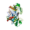
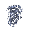
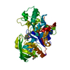
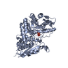
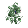
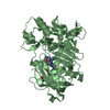
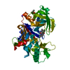
| Title | Crystal structure of aminoglycoside 6'-N-acetyltransferase complexed with coenzyme A | ||||||
 Components Components | aac(6')-Ii | ||||||
 Keywords Keywords | TRANSFERASE / alpha beta protein / N-acetyl transferase | ||||||
| Function / homology |  Function and homology information Function and homology informationacyltransferase activity, transferring groups other than amino-acyl groups Similarity search - Function | ||||||
| Biological species |  Enterococcus faecium (bacteria) Enterococcus faecium (bacteria) | ||||||
| Method |  X-RAY DIFFRACTION / SYNCHROTRON / MOLECULAR REPLACEMENT / Resolution: 2.2 Å X-RAY DIFFRACTION / SYNCHROTRON / MOLECULAR REPLACEMENT / Resolution: 2.2 Å | ||||||
 Authors Authors | Burk, D.L. / Xiong, B. / Breitbach, C. / Berghuis, A.M. | ||||||
 Citation Citation | Journal: Acta Crystallogr.,Sect.D / Year: 2005 Title: Structures of aminoglycoside acetyltransferase AAC(6')-Ii in a novel crystal form: structural and normal-mode analyses. Authors: Burk, D.L. / Xiong, B. / Breitbach, C. / Berghuis, A.M. #1: Journal: Protein Sci. / Year: 2003Title: Crystal Structure Of Aminoglycoside 6'-Acetyltransferase Type II In Complex With Coenzyme A Authors: Burk, D.L. / Ghuman, N. / Wybenga-Groot, L.E. / Berghuis, A.M. #2: Journal: Structure / Year: 1999Title: Crystal structure of an aminoglycoside 6'-N-acetyltransferase: defining the GCN5-related N-acetyltransferase superfamily fold Authors: Wybenga-Groot, L.E. / Draker, K. / Wright, G.D. / Berghuis, A.M. | ||||||
| History |
| ||||||
| Remark 999 | SEQUENCE THE AUTHOR STATES THAT THERE IS AN ERROR IN THE SEQUENCE DATABASE. |
- Structure visualization
| Structure viewer | Molecule: MolmilJmol/JSmol |
|---|
- Downloads & links
-Download
| PDBx/mmCIF format | 2a4n.cif.gz | 94.1 KB | Display | PDBx/mmCIF format |
|---|---|---|---|---|
| PDB format | pdb2a4n.ent.gz | 70.7 KB | Display | PDB format |
| PDBx/mmJSON format | 2a4n.json.gz | Tree view | PDBx/mmJSON format | |
| Others |  Other downloads Other downloads |
-Validation report
| Arichive directory | https://data.pdbj.org/pub/pdb/validation_reports/a4/2a4nftp://data.pdbj.org/pub/pdb/validation_reports/a4/2a4n | HTTPS FTP |
|---|
-Related structure data
| Related structure data |  1n71S S: Starting model for refinement |
|---|---|
| Similar structure data |


-Links
 PDBj
PDBj


- Assembly
| Deposited unit | 
| |||||||||
|---|---|---|---|---|---|---|---|---|---|---|
| 1 |
| |||||||||
| 2 | 
| |||||||||
| Unit cell |
| |||||||||
| Components on special symmetry positions |
| |||||||||
| Details | The asymmetric unit contains the biological unit (dimer). |
-Components
| #1: Protein | Mass: 20730.195 Da / Num. of mol.: 2 Source method: isolated from a genetically manipulated source Source: (gene. exp.) Enterococcus faecium (bacteria) / Gene: aac(6')-Ii / Plasmid: pPLaac-1 / Species (production host): Escherichia coliProduction host: Strain (production host): W3110 References: UniProt: Q47764, aminoglycoside 6'-N-acetyltransferase #2: Chemical |   Mass: 767.534 Da / Num. of mol.: 2 / Source method: obtained synthetically / Formula: C21H36N7O16P3S Mass: 767.534 Da / Num. of mol.: 2 / Source method: obtained synthetically / Formula: C21H36N7O16P3S#3: Chemical |   Mass: 96.063 Da / Num. of mol.: 2 / Source method: obtained synthetically / Formula: SO4 Mass: 96.063 Da / Num. of mol.: 2 / Source method: obtained synthetically / Formula: SO4#4: Water | ChemComp-HOH / |  Mass: 18.015 Da / Num. of mol.: 198 / Source method: isolated from a natural source / Formula: H2O Mass: 18.015 Da / Num. of mol.: 198 / Source method: isolated from a natural source / Formula: H2O |
|---|
-Experimental details
-Experiment
| Experiment | Method: X-RAY DIFFRACTION / Number of used crystals: 1 |
|---|
- Sample preparation
| Crystal | Density Matthews: 2.114 Å3/Da / Density % sol: 40 % |
|---|---|
| Crystal grow | Temperature: 295 K / Method: vapor diffusion, hanging drop / pH: 7.5 Details: HEPES, EDTA, coenzyme A, 2-(6'-N-sisomycin)acetic acid, tri-sodium citrate, ammonium sulfate, pH 7.5, VAPOR DIFFUSION, HANGING DROP, temperature 295K |
-Data collection
| Diffraction | Mean temperature: 95 K |
|---|---|
| Diffraction source | Source: SYNCHROTRON / Site: NSLS  / Beamline: X8C / Wavelength: 1.072 Å / Beamline: X8C / Wavelength: 1.072 Å |
| Detector | Type: ADSC QUANTUM 4 / Detector: CCD / Date: Nov 6, 2002 / Details: mirrors |
| Radiation | Monochromator: mirrors / Protocol: SINGLE WAVELENGTH / Monochromatic (M) / Laue (L): M / Scattering type: x-ray |
| Radiation wavelength | Wavelength: 1.072 Å / Relative weight: 1 |
| Reflection | Resolution: 2.2→100 Å / Num. all: 18341 / Num. obs: 18341 / % possible obs: 99 % / Observed criterion σ(F): 0 / Observed criterion σ(I): 0 / Biso Wilson estimate: 14.6 Å2 / Rmerge(I) obs: 0.076 / Χ2: 1.019 |
| Reflection shell | Resolution: 2.2→2.28 Å / % possible obs: 91.2 % / Rmerge(I) obs: 0.233 / Num. measured obs: 1663 / Χ2: 0.553 / % possible all: 95.4 |
-Phasing
| Phasing MR | Method rotation: fast direct / Method translation: &STRIP%trans_method |
|---|
- Processing
| Software |
| |||||||||||||||||||||||||
|---|---|---|---|---|---|---|---|---|---|---|---|---|---|---|---|---|---|---|---|---|---|---|---|---|---|---|
| Refinement | Method to determine structure: MOLECULAR REPLACEMENT Starting model: PDB entry 1N71 Resolution: 2.2→48.43 Å / Rfactor Rfree error: 0.006 / Data cutoff high absF: 365371.719 / Data cutoff low absF: 0 / Isotropic thermal model: RESTRAINED / Cross valid method: THROUGHOUT / σ(F): 0 / Stereochemistry target values: Engh & Huber
| |||||||||||||||||||||||||
| Solvent computation | Solvent model: FLAT MODEL / Bsol: 55.169 Å2 / ksol: 0.35 e/Å3 | |||||||||||||||||||||||||
| Displacement parameters | Biso mean: 28 Å2
| |||||||||||||||||||||||||
| Refine analyze |
| |||||||||||||||||||||||||
| Refinement step | Cycle: LAST / Resolution: 2.2→48.43 Å
| |||||||||||||||||||||||||
| Refine LS restraints |
| |||||||||||||||||||||||||
| Refine LS restraints NCS | NCS model details: RESTRAINTS | |||||||||||||||||||||||||
| LS refinement shell | Resolution: 2.2→2.34 Å / Rfactor Rfree error: 0.017 / Total num. of bins used: 6
| |||||||||||||||||||||||||
| Xplor file |
|
